



Mutations in the E3 ubiquitin ligase Parkin gene have been linked to early onset Parkinson’s disease. Besides many other roles, Parkin is involved in clearance of damaged mitochondria via mitophagy—a process of particular importance in dopaminergic neurons. Upon mitochondrial damage, Parkin accumulates at the outer mitochondrial membrane and is activated, leading to ubiquitination of many mitochondrial substrates and recruitment of mitophagy effectors. While the activation mechanisms of autoinhibited Parkin have been extensively studied, it remains unknown how Parkin recognizes its substrates for ubiquitination. Here, we characterize a conserved region in the flexible linker between the Ubl and RING0 domains of Parkin, which is indispensable for Parkin interaction with the mitochondrial GTPase Miro1. Our results may explain fast kinetics of Miro1 ubiquitination by Parkin in recombinant assays and provide a biochemical explanation for Miro1-dependent Parkin recruitment to the mitochondrial membrane observed in cells. Our findings are important for understanding mitochondrial homeostasis and may inspire new therapeutic avenues for Parkinson’s disease.
Introduction
Heritable forms of Parkinson’s disease (PD) account for 5–10% of all PD cases. The implication of mutations in PRKN gene, encoding for the E3 ubiquitin ligase Parkin (Shimura et al., 2000; Zhang et al., 2000), in early onset, autosomal recessive parkinsonism was described in 1998 (Kitada et al., 1998). Since then, intensive research efforts have been made to understand Parkin function and structure. With an increasing number of identified PD patient mutations throughout the PRKN sequence (Zhang et al., 2021; Li et al., 2021; Jiang et al., 2020; Kasten et al., 2018; Taghavi et al., 2018), it became clear that all domains, and even supposedly disordered regions of Parkin, are important for its function. Parkin is a RING-BetweenRING-RING E3 ubiquitin ligase (Wenzel et al., 2011), composed of five domains (Riley et al., 2013): an N-terminal ubiquitin-like domain (Ubl), connected by a 65-residue flexible region (linker) to a unique Parkin RING0 domain, which, together with RING1 and BRcat (IBR), constitutes a more rigid “core” of Parkin (Fig. 1). At the C terminus, another partially disordered “tether” region connects the core to the catalytic Rcat domain of Parkin, containing the active site. Several elements in Parkin maintain its closed, autoinhibited conformation: Ubl blocks the predicted donor ubiquitin-binding site, the repressive element of Parkin in the tether region blocks the incoming E2 enzyme–binding site, and RING0 partially occludes the active site cysteine in Rcat (Kumar et al., 2015; Kumar et al., 2017; Chaugule et al., 2011; Trempe et al., 2013; Riley et al., 2013; Wauer et al., 2015). Parkin, like other E3s, catalyzes the last step in the ubiquitination reaction and modifies target proteins on lysine residues with a small protein, ubiquitin. To achieve this, Parkin, as an RING-BetweenRING-RING ligase, is thought to interact with an E2 enzyme charged with ubiquitin, accept ubiquitin onto its active site, interact with or position itself in sufficient proximity to a substrate, and finally transfer ubiquitin onto a target lysine. To fulfil its role, Parkin must first be released from its autoinhibition.

Schematic of Parkin in its autoinhibited conformation as predicted by AlphaFold. Outlined are the structural elements in sequence order: Ubl (blue), linker (purple), RING0, RING1, BRcat (IBR) (green), tether (red), and Rcat (orange). The prediction allows us to appreciate the length of the 65-residue-long linker as compared with Parkin domains.
Schematic of Parkin in its autoinhibited conformation as predicted by AlphaFold. Outlined are the structural elements in sequence order: Ubl (blue), linker (purple), RING0, RING1, BRcat (IBR) (green), tether (red), and Rcat (orange). The prediction allows us to appreciate the length of the 65-residue-long linker as compared with Parkin domains.
The most studied instance for Parkin activation occurs at damaged mitochondria, where activation of Parkin leads to mitophagy (Narendra et al., 2008; Matsuda et al., 2010). Upon induction of damage, the kinase PTEN-induced kinase 1 (PINK1) is stabilized on the outer mitochondrial membrane (OMM) and phosphorylates its primary substrate, ubiquitin, on Ser65 (Kazlauskaite et al., 2014b; Koyano et al., 2014; Kane et al., 2014). Phosphorylated ubiquitin (pUb) is understood to serve for recruitment of Parkin (Okatsu et al., 2015), which is normally localized in the cytoplasm but exhibits nanomolar affinity toward pUb (Kazlauskaite et al., 2015; Ordureau et al., 2014; Kumar et al., 2015). Moreover, PINK1 can also phosphorylate the Ubl of Parkin, especially when it is already released from its autoinhibitory site by pUb binding, which leads to further Parkin activation (Kazlauskaite et al., 2015; Gladkova et al., 2018; Sauvé et al., 2018; Kondapalli et al., 2012; Shiba-Fukushima et al., 2012).
Active Parkin proceeds to ubiquitinate mitochondrial proteins, and the resulting ubiquitin chains constitute a signal for mitophagy (Yamano et al., 2016). Several cellular studies have suggested the existence of dozens of Parkin substrates and hundreds of target lysines (Ordureau et al., 2015; Antico et al., 2021; Sarraf et al., 2013; Chan et al., 2011; Okatsu et al., 2012; Ordureau et al., 2018; Ordureau et al., 2020). These include mitochondrial, but also cytoplasmic and even nuclear proteins. The vast number of potential substrates has led to suggestions that Parkin acts in a promiscuous manner when it is brought into proximity to substrates by the presence of pUb (Okatsu et al., 2015; Koyano et al., 2019b; Dunkerley et al., 2022; Vranas et al., 2022). However, biochemical assays show that Parkin in its active, phosphorylated form (pParkin) ubiquitinates proteins without pUb (Kazlauskaite et al., 2014b; Klosowiak et al., 2016), which suggests direct interactions between pParkin and its substrates likely exist. An alternative hypothesis is that Parkin interacts with substrates containing ubiquitin-interacting motifs via its Ubl domain (Fallon et al., 2006; Chaugule et al., 2011; Spratt et al., 2013). However, most of the reported Parkin substrates have no ubiquitin-interacting motifs.
It has been proposed that the lysines targeted by Parkin are not random and are conserved, although no consensus ubiquitination motif has been found (Klosowiak et al., 2016; Koyano et al., 2019b). While attempts have been made to identify “preferred” Parkin targets, there is no agreement on which mitochondrial substrate is favored by Parkin upon mitochondrial disruption, with mitofusins 1 and 2 (Mfn1/2) (Vranas et al., 2022) or voltage-dependent anion-selective channel proteins proposed (Ordureau et al., 2018; Narendra and Youle, 2024). Interestingly, a recent study reported that Mfn1/2 and the mitochondrial GTPase Miro1 are the only detectable OMM proteins whose abundance is decreased upon mitochondria depolarization in a p97-dependent fashion (Ordureau et al., 2020), which may indicate some level of Parkin specificity toward Mfn1/2 and Miro1. Biochemically, Parkin does exhibit preference toward the C-terminal GTPase domain of Miro1 (Klosowiak et al., 2016; Dunkerley et al., 2022), as compared with its close homolog, Miro2. Mitochondrial Rho GTPase (Miro) family of proteins consists of two GTPase domains flanking two EF-hand domains and a C-terminal transmembrane helix that anchors Miro onto the OMM. Miro proteins are involved in mitochondrial contacts with other organelles, such as peroxisomes and endoplasmic reticulum, and with the cytoskeleton. Miro proteins have been shown to regulate mitochondrial shape dynamics in mitophagy and in mitochondrial quality control, which, when perturbed, can lead to neurodegeneration (reviewed in Eberhardt et al. [2020]). Recently, Miro1 mutations have been identified in patients with PD (Berenguer-Escuder et al., 2019; Berenguer-Escuder et al., 2020; Grossmann et al., 2019). The key biochemical difference between Miro1 and Miro2 resides in the presence of a specific lysine K572 in Miro1, which has a particular chemical environment, favorable for ubiquitin conjugation by Parkin (Klosowiak et al., 2016). Importantly, it has been suggested that Parkin acquires specificity when it is phosphorylated, while artificially activated Parkin (for example, containing an N-terminal tag [Burchell et al., 2012]) acts indiscriminately (Klosowiak et al., 2016). While the molecular basis for preferential ubiquitination of K572 has been described from the Miro1 perspective, there is no biochemical evidence for Parkin interaction with Miro1 or any other substrate, although recently an interface between Miro1 and Parkin has been proposed based on AlphaFold prediction (Covill-Cooke et al., 2024). It is also unclear whether Parkin can indeed directly ubiquitinate all the suggested proteins in its native, non-tagged form.
Here, we evaluate a set of different proteins as Parkin substrates in biochemical assays and show that untagged, full-length Parkin activated with phosphorylation can ubiquitinate many lysine-containing proteins. Interestingly, we observe that Miro1 is ubiquitinated most efficiently by active Parkin in a reconstituted in vitro assay. Based on this observation, we focus on Parkin interaction with Miro1 and establish an assay to stabilize the Parkin–Miro1 complex. We then characterize the stabilized complex using a series of biochemical and biophysical methods. Most importantly, we describe a substrate-interacting site in Parkin located in the disordered linker region between Ubl and RING0 domains of Parkin. Upon disruption of Parkin–Miro1 interaction by competition or amino acid substitution, Miro1 ubiquitination by Parkin is compromised. Our findings uncover a substrate recognition mechanism in Parkin and explain previous observations in cells (Safiulina et al., 2018; Covill-Cooke et al., 2024).
Results
Active Parkin efficiently ubiquitinates a variety of proteins in vitro
Studies report a myriad of potential Parkin substrates located throughout the cell, including cytoplasmic, mitochondrial, and nuclear proteins (Ordureau et al., 2015; Antico et al., 2021; Sarraf et al., 2013; Chan et al., 2011; Okatsu et al., 2012). However, these studies often use tagged versions of Parkin, which are known to result in artificial Parkin activation (Burchell et al., 2012; Matsuda et al., 2006; Chaugule et al., 2011) and loss of specificity (Klosowiak et al., 2016). We therefore wanted to evaluate the promiscuity of untagged, full-length Parkin in biochemical assays. For this, we first looked at ubiquitination of a selected set of three confirmed mitochondrial substrates (Gegg et al., 2010; Tanaka et al., 2010; Kazlauskaite et al., 2014a; Wang et al., 2011; Narendra et al., 2012; Okatsu et al., 2012; Chan et al., 2011; Narendra and Youle, 2024) as constructs which are suitable for biochemical studies: Miro1 (HA-tagged, 181–579) (Klosowiak et al., 2016), Mfn1 (6His-tagged, 1–364-GSGSGSGGS-694–741) (Yan et al., 2018), and mitoNEET/CISD1 (6His-tagged, 33–108) (Conlan et al., 2009) (Fig. S1). We have chosen these particular constructs of Miro1, Mfn1, and mitoNEET because they have previously been reported as amenable for purification in high amounts and high purity, as required for our reconstituted in vitro assays, unlike the full-length proteins. As previously established, to be active for ubiquitination, Parkin requires the allosteric activator pUb, or phosphorylation at Ser65 in the Ubl of Parkin, or both (Kazlauskaite et al., 2014b; Shiba-Fukushima et al., 2012; Koyano et al., 2014; Kane et al., 2014). It has been proposed that Parkin is brought in proximity to its targets via pUb on the mitochondrial membrane (Okatsu et al., 2015; Koyano et al., 2019b; Dunkerley et al., 2022; Vranas et al., 2022). However, in our assays, we observe that pParkin can ubiquitinate substrates without pUb (Fig. S1), consistent with observations by other groups (Kazlauskaite et al., 2014a; Klosowiak et al., 2016). This suggests that alternate interactions exist between pParkin and its substrates in addition to those driven by pUb recruitment.
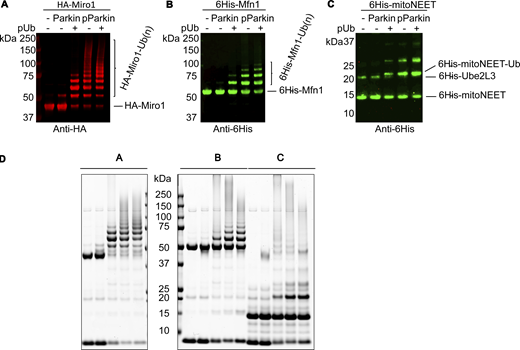
Ubiquitination assays of Parkin mitochondrial substrates. (A–C) Substrates Miro1 (A), Mfn1 (B), and mitoNEET (C) were submitted to Parkin ubiquitination assays in the presence or absence of pUb and with Parkin or pParkin, as indicated. Reactions were resolved by SDS-PAGE and revealed with western blotting as indicated. (D) Corresponding Coomassie-stained gels. Source data are available for this figure: SourceData FS1.
Ubiquitination assays of Parkin mitochondrial substrates. (A–C) Substrates Miro1 (A), Mfn1 (B), and mitoNEET (C) were submitted to Parkin ubiquitination assays in the presence or absence of pUb and with Parkin or pParkin, as indicated. Reactions were resolved by SDS-PAGE and revealed with western blotting as indicated. (D) Corresponding Coomassie-stained gels. Source data are available for this figure: SourceData FS1.
Therefore, to simplify our assay setup and focus on direct Parkin–substrate interactions, we performed subsequent assays with pParkin in the absence of pUb. We assessed pParkin activity toward Miro1, Mfn1, and mitoNEET constructs, as well as pParkin itself, ubiquitin, and typical components of ubiquitination assays, such as the E2 enzyme UBE2L3 and the E1 enzyme UBE1, in a time-resolved assay (Fig. 2, A and B). All tested proteins are modified with ubiquitin by pParkin. In addition, we tested a nuclear protein, FANCD2, not reported as a Parkin substrate in any of the extensive substrate lists nor as Parkin interactors in the BioGRID database (Oughtred et al., 2021). Despite not being a known substrate, FANCD2 is also ubiquitinated (Fig. 2, A and B). Finally, we tested a short linear peptide containing three lysine residues and found that it is also modified by pParkin in vitro (Fig. 2, A and B). Our data confirm that Parkin in its active conformation can modify many proteins that contain solvent-exposed lysines.
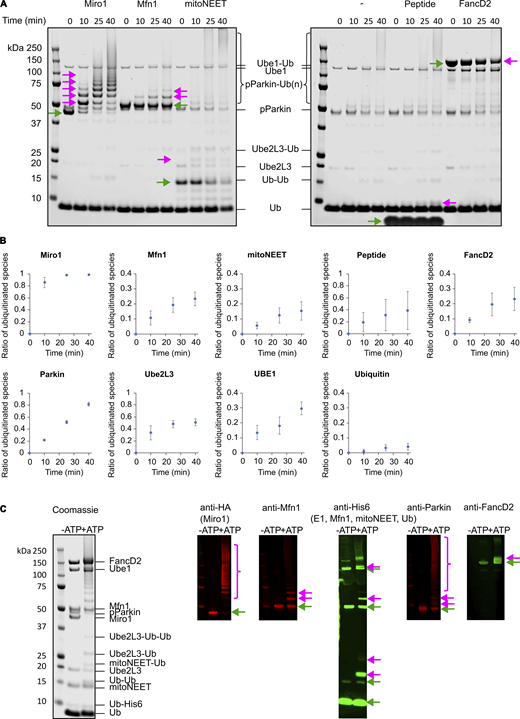
Active Parkin ubiquitinates various proteins but most efficiently polyubiquitinates Miro1. (A) Time-resolved ubiquitination assays were performed with pParkin and various proteins as substrates as indicated and resolved on SDS-PAGE. Represented are Coomassie-stained gels. Green arrows indicate the unmodified substrates, and pink arrows indicate the ubiquitinated substrates. (B) Quantification of ubiquitinated species generated by pParkin over time from at least three replicate experiments. Error bars represent standard deviations. (C) Active Parkin ubiquitinates all proteins present in the assay and detectably polyubiquitinates Miro1 and itself. Miro1, mitoNEET, Mfn1, and FancD2 were added to a ubiquitination mix containing ubiquitin (Ub), Ube1 (E1), Ube2L3, and pParkin. Ubiquitinated species were detected using Coomassie and western blotting as indicated. Source data are available for this figure: SourceData F2.
Active Parkin ubiquitinates various proteins but most efficiently polyubiquitinates Miro1. (A) Time-resolved ubiquitination assays were performed with pParkin and various proteins as substrates as indicated and resolved on SDS-PAGE. Represented are Coomassie-stained gels. Green arrows indicate the unmodified substrates, and pink arrows indicate the ubiquitinated substrates. (B) Quantification of ubiquitinated species generated by pParkin over time from at least three replicate experiments. Error bars represent standard deviations. (C) Active Parkin ubiquitinates all proteins present in the assay and detectably polyubiquitinates Miro1 and itself. Miro1, mitoNEET, Mfn1, and FancD2 were added to a ubiquitination mix containing ubiquitin (Ub), Ube1 (E1), Ube2L3, and pParkin. Ubiquitinated species were detected using Coomassie and western blotting as indicated. Source data are available for this figure: SourceData F2.
Miro1 is ubiquitinated more efficiently than other pParkin’s substrates in vitro
In our assay, pParkin appears to ubiquitinate Miro1 much quicker and more extensively than any other tested protein (Fig. 2, A and B), consistent with reports suggesting Miro1 as the preferred Parkin target (Klosowiak et al., 2016; Dunkerley et al., 2022; Chan et al., 2011). In contrast to the other tested substrates, nearly all Miro1 is ubiquitinated after 40 min of reaction under the conditions used (Fig. 2, A and B).
We then asked whether pParkin would show the same activity toward Miro1 and other substrates in a reaction with a mix of substrates, or whether the presence of Miro1 increases pParkin processivity toward substrates in general. For this, we set up an assay with various Parkin substrates present in the same reaction: Miro1, Mfn1, mitoNEET, non-conjugatable Ub-6His, and FANCD2. We observe that pParkin ubiquitinates each tested protein to similar levels as in reactions with each substrate separately (Fig. 2 C), with Miro1 and pParkin ubiquitinated much more extensively as compared with other proteins. Given this observation, we quantified pParkin efficiency in processing ubiquitin, when presented with each substrate, by measuring the remaining unconjugated ubiquitin in reactions with individual substrates (Fig. 3, A and B). Our data show that pParkin is the most efficient in conjugating ubiquitin to Miro1.
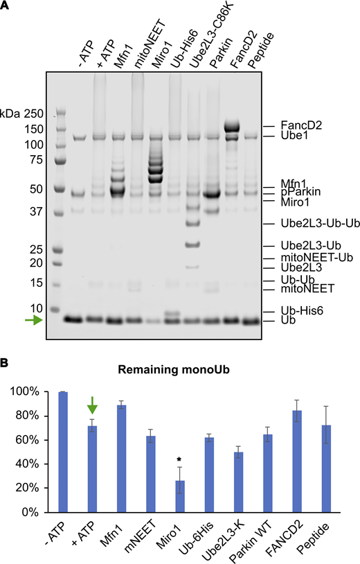
Active Parkin processes ubiquitin most efficiently with Miro1 as substrate. (A) Ubiquitination assays were performed with various substrates, resolved with SDS-PAGE, and visualized by Coomassie staining. Green arrow indicates the band corresponding to unconjugated ubiquitin. (B) Consumption of ubiquitin was assessed by measuring the remaining unconjugated ubiquitin for each substrate using band densitometry from at least three experiments. Error bars represent standard deviations estimated from at least three replicates. Green arrow indicates the positive control used to calculate significance. The significance of the ubiquitin consumption decrease was evaluated using two-sided t test between the +ATP control and the reaction with each substrate, and the P value was below 0.05 only for Miro1, indicated with asterisk. Source data are available for this figure: SourceData F3.
Active Parkin processes ubiquitin most efficiently with Miro1 as substrate. (A) Ubiquitination assays were performed with various substrates, resolved with SDS-PAGE, and visualized by Coomassie staining. Green arrow indicates the band corresponding to unconjugated ubiquitin. (B) Consumption of ubiquitin was assessed by measuring the remaining unconjugated ubiquitin for each substrate using band densitometry from at least three experiments. Error bars represent standard deviations estimated from at least three replicates. Green arrow indicates the positive control used to calculate significance. The significance of the ubiquitin consumption decrease was evaluated using two-sided t test between the +ATP control and the reaction with each substrate, and the P value was below 0.05 only for Miro1, indicated with asterisk. Source data are available for this figure: SourceData F3.
Taken together, these data indicate that while pParkin ubiquitinates various proteins in an in vitro assay, its processivity is increased toward our Miro1 construct as substrate. These results suggest that pParkin’s affinity toward Miro1 may be higher than toward other tested substrates.
Parkin interacts with Miro1 with low affinity
Despite the vast evidence of efficient ubiquitination of various proteins by Parkin, and although many papers report interaction between Parkin and Miro1 (López-Doménech et al., 2021; Safiulina et al., 2018; Covill-Cooke et al., 2024; Shlevkov et al., 2016), there are no reports of quantifiable, direct interaction between Parkin and any of its targets. However, since our assays are performed in the absence of pUb, we assume pParkin must interact with its substrates directly, likely through transient, low-affinity interactions. To capture a Parkin–substrate complex, we chose to investigate a hypothetical interaction with Miro1, based on the observed pParkin’s increased activity toward Miro1 in our in vitro assays. Indeed, it has already been suggested that Parkin interacts transiently with Miro1 in cells even in the absence of mitochondrial damage (Safiulina et al., 2018), and a predicted interaction has been recently reported (Covill-Cooke et al., 2024). To capture the Parkin–Miro1 complex, we tried several methods; however, we were not able to observe a stable complex. In size-exclusion chromatography (SEC), Parkin and Miro1 elute as separate peaks (Fig. S2 A). On native PAGE, we do not observe any additional bands as compared with controls (Fig. S2 B). Isothermal titration calorimetry does not detect any heat exchange when Miro1 is titrated into Parkin (Fig. S2 C) (Dunkerley et al., 2022). Surface plasmon resonance does not detect any interaction either (Fig. S2 D). These data show that the Parkin–Miro1 interaction is weak and cannot be detected by these biophysical methods.
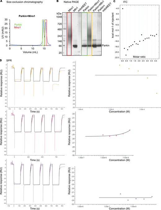
Results from attempts using various methods to capture Parkin–Miro1 complex. (A–D) A: SEC, B: native PAGE, C: ITC, and D: SPR. mAU: mili-arbitrary units; RU: response units. Source data are available for this figure: SourceData FS2. ITC, isothermal titration calorimetry; SPR, surface plasmon resonance.
Results from attempts using various methods to capture Parkin–Miro1 complex. (A–D) A: SEC, B: native PAGE, C: ITC, and D: SPR. mAU: mili-arbitrary units; RU: response units. Source data are available for this figure: SourceData FS2. ITC, isothermal titration calorimetry; SPR, surface plasmon resonance.
Parkin–Miro1 complex is detectable upon chemical cross-linking
As we could not detect a stable complex of Parkin and Miro1 through biophysical methods, we wanted to explore whether chemical cross-linking could stabilize the interaction. For this, we used a disuccinimidyl suberate (DSS) cross-linker, which couples primary amines with a spacer of 11.4 Å. After optimization, we detect a prominent band corresponding to non-phosphorylated Parkin–Miro1 complex, as confirmed by Coomassie staining (Fig. 4 A) and western blot analysis (Fig. 4 B). Importantly, a control Parkin-Mfn1 cross-linking reaction produces only a very faint band corresponding to a potential complex, as compared with Parkin–Miro1 cross-linked complex. Our Mfn1 construct has 19 surface-exposed lysines (Fig. 4 C); therefore, the lack of cross-linking is not due to the unavailability of lysine residues. In contrast, the Parkin-Miro1 cross-linking is easily detectable, indicating a more favorable interaction. The position of the 100-kDa band on SDS-PAGE, compared with the Parkin and Miro1 bands, suggests a 1:1 stoichiometry (Fig. 4 A).
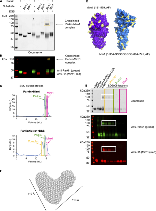
Capture of Parkin–Miro1 complex with a cross-linking assay, isolation, and characterization by SAXS. (A) Parkin was incubated with Miro1 or a negative control Mfn1, and the formed complexes were stabilized in a cross-linking reaction by adding DSS where indicated. Controls include each protein separately with and without DSS. (B) After quenching the cross-linking reaction, the samples were resolved by SDS-PAGE and stained with Coomassie or by western blotting (WB) with anti-Parkin (green) and anti-HA (Miro1) (red) antibodies. The band corresponding to the cross-linked 1:1 Parkin–Miro1 complex is indicated with an arrow and yellow box. (C) AlphaFold-generated surface representation of Miro1 and Mfn1 constructs used in the assay, with exposed lysines colored in magenta. (D) SEC of cross-linked Parkin–Miro1 complex. Graphs represent elution profiles of the samples without (top) and with the DSS cross-linker (bottom). The third peak corresponding to the complex appears in the SEC run with the cross-linker (marked with a yellow box). (E) SEC fractions were run on SDS-PAGE and revealed with Coomassie staining or by WB. (F) Ab initio SAXS model of the cross-linked Parkin–Miro1 complex. Source data are available for this figure: SourceData F4.
Capture of Parkin–Miro1 complex with a cross-linking assay, isolation, and characterization by SAXS. (A) Parkin was incubated with Miro1 or a negative control Mfn1, and the formed complexes were stabilized in a cross-linking reaction by adding DSS where indicated. Controls include each protein separately with and without DSS. (B) After quenching the cross-linking reaction, the samples were resolved by SDS-PAGE and stained with Coomassie or by western blotting (WB) with anti-Parkin (green) and anti-HA (Miro1) (red) antibodies. The band corresponding to the cross-linked 1:1 Parkin–Miro1 complex is indicated with an arrow and yellow box. (C) AlphaFold-generated surface representation of Miro1 and Mfn1 constructs used in the assay, with exposed lysines colored in magenta. (D) SEC of cross-linked Parkin–Miro1 complex. Graphs represent elution profiles of the samples without (top) and with the DSS cross-linker (bottom). The third peak corresponding to the complex appears in the SEC run with the cross-linker (marked with a yellow box). (E) SEC fractions were run on SDS-PAGE and revealed with Coomassie staining or by WB. (F) Ab initio SAXS model of the cross-linked Parkin–Miro1 complex. Source data are available for this figure: SourceData F4.
To gain more insight into the size and shape of the Parkin–Miro1 complex in solution, we isolated the cross-linked complex using SEC and submitted it to SEC-SAXS (small-angle scattering) analysis (Figs. 2, D–F and S3). As controls, we also analyzed Parkin and Miro1 separately. The estimated radius of gyration for the complex is 39 Å, as compared with 28 Å and 28.5 Å determined for autoinhibited Parkin and Miro1 alone, respectively (Fig. S3). The radius of gyration values we obtained for Parkin and Miro1 are consistent with the values published previously (Klosowiak et al., 2013; Spratt et al., 2013). An ab initio model of the Parkin–Miro1 complex suggests a V-shaped conformation, with the length of the branches estimated at around 110 Å and 116 Å (Fig. 4 F), likely corresponding to the two cross-linked proteins linked near their termini, with the other extremities pointing outward. These data indicate an interaction between Parkin and Miro1 that is stabilized by chemical cross-linking.

SEC-SAXS analysis of cross-linked Parkin–Miro1 complex in comparison with Parkin and Miro1 alone. (A) Experimental intensity plots of Parkin (green), Miro1 (purple), and cross-linked Parkin–Miro1 complex (orange). (B and C) Guinier plot of Parkin, Miro1, and cross-linked Parkin–Miro1 complex (B) with residuals shown in C. Rg values of 28 Å for Parkin, 28.5 Å for Miro1, and 39 Å for the complex were derived from these data. (D) Overlay of the SAXS ab initio model with an AlphaFold-predicted Parkin–Miro1 complex. Rg: radius of gyration; ATSAS: software suite for data analysis of small-angle scattering (SAS).
SEC-SAXS analysis of cross-linked Parkin–Miro1 complex in comparison with Parkin and Miro1 alone. (A) Experimental intensity plots of Parkin (green), Miro1 (purple), and cross-linked Parkin–Miro1 complex (orange). (B and C) Guinier plot of Parkin, Miro1, and cross-linked Parkin–Miro1 complex (B) with residuals shown in C. Rg values of 28 Å for Parkin, 28.5 Å for Miro1, and 39 Å for the complex were derived from these data. (D) Overlay of the SAXS ab initio model with an AlphaFold-predicted Parkin–Miro1 complex. Rg: radius of gyration; ATSAS: software suite for data analysis of small-angle scattering (SAS).
Identification of Parkin–Miro1 interaction site
After establishing the cross-linking assay to capture a Parkin–Miro1 complex, we sought to identify the regions of Parkin that are important for the interaction with Miro1. For this, we performed cross-linking reactions with various Parkin deletion constructs, devoid of Ubl, linker, Ubl and linker, or Rcat (Fig. 5 A). Interestingly, only the constructs without a linker exhibit decreased cross-linking efficiency with Miro1, suggesting that the linker element of Parkin is involved in the interaction. Of note, we have previously observed that Parkin’s ability to ubiquitinate Miro1 is compromised when the linker region is deleted (Kumar et al., 2015). No decrease in complex formation is observed with the deltaRcat variant, which suggests the interaction of Rcat with the substrate, although likely necessary for the ubiquitination activity (Klosowiak et al., 2016), does not contribute to the overall stability of the complex. For confirmation, we tested a Parkin construct composed of Ubl-linker (residues 1–143) and observed that this region of Parkin is sufficient for cross-linking (Fig. 5 A). Interestingly, when we perform the cross-linking reaction with phosphorylated Parkin, we do not observe increased intensity of the band corresponding to the cross-linked complex. This indicates that cross-linking of Parkin and Miro1 is not dependent on the activation state of Parkin, consistent with previous cellular observations (Safiulina et al., 2018).
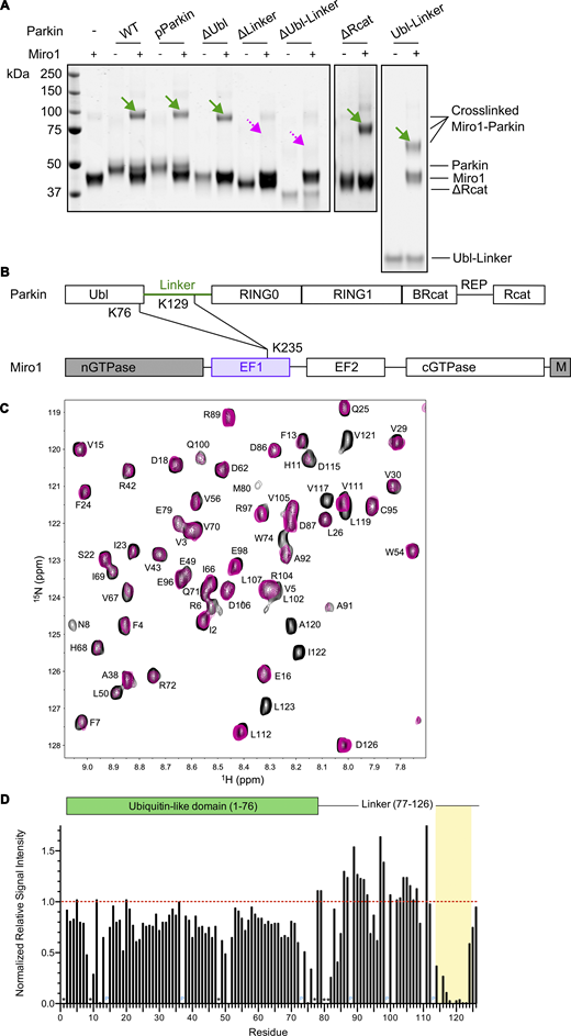
Cross-linking assay, mass spectrometry, and NMR spectroscopy experiments define the Miro1-interacting region in Parkin. (A) Series of Parkin mutants with various domains deleted were assayed in the cross-linking assay for their ability to form a cross-linkable complex with Miro1. Green arrows indicate bands corresponding to cross-linked complex, while dashed pink arrows point to areas where bands for cross-linked complex were not detected. (B) Schematic of Parkin and Miro1 domains with indicated cross-linked lysines as identified by mass spectrometry. Parkin construct is full-length, while the Miro1 construct (residues 181–579) does not have the N-terminal GTPase domain nor the C-terminal transmembrane domain (grey). (C) Interaction of Ubl1–126 and Miro1180–582 using NMR spectroscopy. Overlay of regions of the 1H-15N HSQC spectra of 15N Ubl1–126 alone (black contours) and 15N-labeled Ubl1–126 with one equivalent of unlabeled Miro1180-582 (pink contours). Residues are labeled according to their one-letter amino acid code and number. G114 and S116 are not shown in this region. (D) Relative signal intensity for each residue was calculated as the ratio of 15N-UBL1–126 signal intensities after the addition of unlabeled Miro1180–582 to those recorded before its addition. Values were normalized to the highest intensity signals observed after substrate addition, specifically in UBL1–126 residues 78–112. Residues without assignments are indicated with an asterisk (*), and proline residues are indicated in blue (P). A dashed line (1.0) represents the intensity ratio for signals unaffected by Miro1 binding, exemplifying the reduction in signal intensity for residues 115–124 in UBL1–126, highlighted in yellow. Source data are available for this figure: SourceData F5.
Cross-linking assay, mass spectrometry, and NMR spectroscopy experiments define the Miro1-interacting region in Parkin. (A) Series of Parkin mutants with various domains deleted were assayed in the cross-linking assay for their ability to form a cross-linkable complex with Miro1. Green arrows indicate bands corresponding to cross-linked complex, while dashed pink arrows point to areas where bands for cross-linked complex were not detected. (B) Schematic of Parkin and Miro1 domains with indicated cross-linked lysines as identified by mass spectrometry. Parkin construct is full-length, while the Miro1 construct (residues 181–579) does not have the N-terminal GTPase domain nor the C-terminal transmembrane domain (grey). (C) Interaction of Ubl1–126 and Miro1180–582 using NMR spectroscopy. Overlay of regions of the 1H-15N HSQC spectra of 15N Ubl1–126 alone (black contours) and 15N-labeled Ubl1–126 with one equivalent of unlabeled Miro1180-582 (pink contours). Residues are labeled according to their one-letter amino acid code and number. G114 and S116 are not shown in this region. (D) Relative signal intensity for each residue was calculated as the ratio of 15N-UBL1–126 signal intensities after the addition of unlabeled Miro1180–582 to those recorded before its addition. Values were normalized to the highest intensity signals observed after substrate addition, specifically in UBL1–126 residues 78–112. Residues without assignments are indicated with an asterisk (*), and proline residues are indicated in blue (P). A dashed line (1.0) represents the intensity ratio for signals unaffected by Miro1 binding, exemplifying the reduction in signal intensity for residues 115–124 in UBL1–126, highlighted in yellow. Source data are available for this figure: SourceData F5.
To investigate the details of the interaction, we analyzed the cross-linked full-length Parkin–Miro1 Coomassie-stained band by mass spectrometry. We find that lysines 76 and 129 in Parkin, located in the C terminus of the Ubl domain and in the linker region, respectively, are uniquely cross-linked to lysine 235 in the EF1 domain in Miro1 (Figs. 5 B, S4 A, S5, S6, and Table S1). This further confirms that the linker region in Parkin is involved in the interaction with Miro1, while the Ubl domain remains in cross-linkable proximity (<30 Å [Merkley et al., 2014]) to the interaction site. We find similar results when cross-linking pParkin with Miro1 (Figs. S4 B, S7, S8, and Table S1), suggesting that the interaction is not affected by the activation state of Parkin.

Identification of cross-linked lysines in Parkin–Miro1 complex by mass spectrometry. (A and B) Unique cross-linking sites and decoy analysis performed using MEROX for (A) Parkin–Miro1 and (B) pParkin–Miro1 complex. FDR, false discovery rate.
Identification of cross-linked lysines in Parkin–Miro1 complex by mass spectrometry. (A and B) Unique cross-linking sites and decoy analysis performed using MEROX for (A) Parkin–Miro1 and (B) pParkin–Miro1 complex. FDR, false discovery rate.

Identification of cross-linked lysines in Parkin–Miro1 complex by mass spectrometry. Spectra and cross-link position for the highest scoring interprotein peptides in Parkin–Miro1 complex.
Identification of cross-linked lysines in Parkin–Miro1 complex by mass spectrometry. Spectra and cross-link position for the highest scoring interprotein peptides in Parkin–Miro1 complex.

Identification of cross-linked lysines in Parkin–Miro1 complex by mass spectrometry. Spectra and cross-link position for the second highest scoring interprotein peptides in Parkin–Miro1 complex.
Identification of cross-linked lysines in Parkin–Miro1 complex by mass spectrometry. Spectra and cross-link position for the second highest scoring interprotein peptides in Parkin–Miro1 complex.

Identification of cross-linked lysines in Parkin–Miro1 complex by mass spectrometry. Spectra and cross-link position for the highest scoring interprotein peptides in pParkin–Miro1 complex.
Identification of cross-linked lysines in Parkin–Miro1 complex by mass spectrometry. Spectra and cross-link position for the highest scoring interprotein peptides in pParkin–Miro1 complex.

Identification of cross-linked lysines in Parkin–Miro1 complex by mass spectrometry. Spectra and cross-link position for the second highest scoring interprotein peptides in pParkin–Miro1 complex.
Identification of cross-linked lysines in Parkin–Miro1 complex by mass spectrometry. Spectra and cross-link position for the second highest scoring interprotein peptides in pParkin–Miro1 complex.
To further explore the Miro1 interaction region in Parkin, we used nuclear magnetic resonance (NMR) spectroscopy. We titrated the EF1-EF2-cGTPase region of Miro1 (Klosowiak et al., 2016) (residues 180–582, Miro1180–582) into an 15N-labelled Ubl-linker construct (Ubl1–126) from Parkin. In the absence of Miro1180–582, the 1H-15N heteronuclear single-quantum coherence (HSQC) spectrum of the Ubl1–126 displays well-resolved signals from both the folded Ubl domain and the linker region (Fig. 5, C and D). Notably, most signals for the linker region between M80-D126 fall within 8.0–8.5 ppm, indicative of a disordered protein structure. Upon addition of Miro1180–582, signals from residues D115–V117 and L119–L123 broaden or shift beyond recognition. This observation is typical of a weaker interaction between the two proteins on the intermediate time scale, in agreement with our biophysical experiments. In contrast, all signals from the folded Ubl domain (I2-V70) retain similar intensities and positions in the spectrum. These observations are consistent with binding of the D115–L123 region in Parkin to the 44-kDa fragment of Miro1, with little or no involvement of the Ubl domain, in agreement with our observations from the cross-linking assay.
AlphaFold prediction of Parkin–Miro1 complex indicates a confident region of interaction
To further characterize the elusive Parkin–Miro1 interaction, we employed AlphaFold using ColabFold (Jumper et al., 2021; Mirdita et al., 2022). Parkin–Miro1 interaction prediction has been recently reported (Covill-Cooke et al., 2024). AlphaFold predicts a model for Parkin–Miro1 complex with low confidence with regard to the relative orientation of Parkin and Miro1 domains, as indicated by the predicted aligned error plot (Fig. 6, A and B). Importantly however, there is one region of Parkin predicted as interacting with high confidence with Miro1. It is positioned within the disordered linker region of Parkin, as identified in our cross-linking mass spectrometry experiments, and, more specifically, it overlaps with the region identified from NMR experiments. It corresponds to residues 115–124 (DSVGLAVILHT) in Parkin, which we named the Miro-targeting region (MTR) (Fig. 6, A–C). The MTR is conserved from fish to humans (Fig. 6 C) and includes the hydrophobic “glycine-leucine-alanine-valine-isoleucine-leucine (GLAVIL) motif” (Fig. 6 C) positioned downstream to the conserved “activating element” (ACT) (Gladkova et al., 2018). The MTR is predicted by AlphaFold to thread through the EF1 domain of Miro1, with the hydrophobic residues in the MTR (V117, L119, A120, V121, I122, and L123) binding to a deep hydrophobic pocket in Miro1 (Fig. 6, F and G). The hydrophobic interactions are gated by a triad of charged residues, D115, D126, and R128, with the potential to form salt bridges with Miro1 residues R263, R261 and D228, respectively (Fig. 6 F). AlphaFold contacts, corresponding to pairs of residues confidently predicted to interact in the two proteins, extend to 15, mostly hydrophobic, residues in the Miro1 pocket, paired with Parkin residues in the 115–124 stretch (Fig. 6, D and E). Alternative AlphaFold models indicate the position of the MTR in Parkin relative to Miro1 remains consistent with high confidence scores for the interaction (Fig. S9, A–C).

High-confidence AlphaFold prediction of Parkin–Miro1 interface, which is driven by hydrophobic interactions. (A) Ribbon representation of top-ranked AlphaFold model of Parkin–Miro1 complex, colored according to model confidence (predicted Local Distance Difference Test [pLDDT]) as indicated in the model confidence legend. (B) Predicted aligned error plot reveals a stretch of residues in Parkin linker predicted with high confidence to bind to Miro1. Green boxes highlight the region of interest. Parkin and Miro1 domains are annotated. (C) Sequence alignment of Parkin linker region (residues E98–S131 in Homo sapiens) reveals two patches of residues with higher conservation: the ACT motif and the Miro1-interacting MTR, encompassing the GLAVIL motif. (D) Close-up on the AlphaFold coevolution contacts between the EF1 domain in Miro1 (purple) and Parkin linker region (residues D115–K129, green). The lysines K129 (Parkin) and K235 (Miro1) that were identified as cross-linked by mass spectrometry and the predicted distance between them are indicated. (E) List of the interacting residue pairs in Miro1 and Parkin as illustrated in D. Hydrophobic residues are highlighted in yellow. (F) Hydrophobic surface representation (yellow: hydrophobic residues, blue: hydrophilic residues) of Parkin-binding pocket in the Miro1 EF1 domain with green ribbon representing the linker region in Parkin (D115–R128), which contains the Miro1-interacting region. Residues of interest are represented as sticks. (G) View of the Miro1 EF1 domain (purple ribbon and surface representation) and Parkin D115–R128 region represented as hydrophobic surface, with key hydrophobic residues in Parkin indicated by arrows.
High-confidence AlphaFold prediction of Parkin–Miro1 interface, which is driven by hydrophobic interactions. (A) Ribbon representation of top-ranked AlphaFold model of Parkin–Miro1 complex, colored according to model confidence (predicted Local Distance Difference Test [pLDDT]) as indicated in the model confidence legend. (B) Predicted aligned error plot reveals a stretch of residues in Parkin linker predicted with high confidence to bind to Miro1. Green boxes highlight the region of interest. Parkin and Miro1 domains are annotated. (C) Sequence alignment of Parkin linker region (residues E98–S131 in Homo sapiens) reveals two patches of residues with higher conservation: the ACT motif and the Miro1-interacting MTR, encompassing the GLAVIL motif. (D) Close-up on the AlphaFold coevolution contacts between the EF1 domain in Miro1 (purple) and Parkin linker region (residues D115–K129, green). The lysines K129 (Parkin) and K235 (Miro1) that were identified as cross-linked by mass spectrometry and the predicted distance between them are indicated. (E) List of the interacting residue pairs in Miro1 and Parkin as illustrated in D. Hydrophobic residues are highlighted in yellow. (F) Hydrophobic surface representation (yellow: hydrophobic residues, blue: hydrophilic residues) of Parkin-binding pocket in the Miro1 EF1 domain with green ribbon representing the linker region in Parkin (D115–R128), which contains the Miro1-interacting region. Residues of interest are represented as sticks. (G) View of the Miro1 EF1 domain (purple ribbon and surface representation) and Parkin D115–R128 region represented as hydrophobic surface, with key hydrophobic residues in Parkin indicated by arrows.

Multiple AlphaFold models predict the same Parkin–Miro1-interacting region with high confidence. (A) Overlay of three AlphaFold models represented as ribbons, aligned to Miro1 structure (purple). (B) Close-up on Parkin–Miro1 interface from A. Miro1 EF1 domain is represented as hydrophobic surface featuring a hydrophobic pocket (yellow). Colored ribbons represent the MTR in Parkin. (C) PAE plots for three AlphaFold models of Parkin–Miro1 interaction. Parkin MTR consistently scores a high confidence score (blue) for its interaction with Miro1. (D) AlphaFold prediction of Parkin interaction with full-length Miro1. Blue: N-terminal GTPase domain absent in our construct; yellow stick: S156 reported to be phosphorylated in cells; red sticks: calcium-binding residues in Miro1. (E) Corresponding PAE plot. (F) AlphaFold3 model of Parkin phosphorylated at Ser65 (pParkin) with Miro1. (G) Corresponding PAE plot. PAE, predicted aligned error.
Multiple AlphaFold models predict the same Parkin–Miro1-interacting region with high confidence. (A) Overlay of three AlphaFold models represented as ribbons, aligned to Miro1 structure (purple). (B) Close-up on Parkin–Miro1 interface from A. Miro1 EF1 domain is represented as hydrophobic surface featuring a hydrophobic pocket (yellow). Colored ribbons represent the MTR in Parkin. (C) PAE plots for three AlphaFold models of Parkin–Miro1 interaction. Parkin MTR consistently scores a high confidence score (blue) for its interaction with Miro1. (D) AlphaFold prediction of Parkin interaction with full-length Miro1. Blue: N-terminal GTPase domain absent in our construct; yellow stick: S156 reported to be phosphorylated in cells; red sticks: calcium-binding residues in Miro1. (E) Corresponding PAE plot. (F) AlphaFold3 model of Parkin phosphorylated at Ser65 (pParkin) with Miro1. (G) Corresponding PAE plot. PAE, predicted aligned error.
Since we used a Miro1 construct that has its N-terminal GTPase domain deleted, we wondered whether full-length Miro1 would be predicted to bind to Parkin differently. However, the AlphaFold model for the full-length Miro1 in complex with Parkin closely resembles the one with our Miro1 construct (Fig. S9, D and E), with the N-terminal domain flexibly connected to the remaining Miro1 core. This prediction suggests that the N-terminal GTPase domain does not contribute to the interaction with Parkin. We also tested AlphaFold3 model of phosphorylated Parkin–Miro1 interaction, which seems to predict the same binding site (Fig. S9, F and G), consistent with our cross-linking data, although this prediction has limitations (Pak et al., 2023; Abramson et al., 2024).
We noticed that in the AlphaFold model, the position of the cross-linked K76 residue in Parkin (Fig. 5 B) is predicted to be over 30 Å away from K235 in Miro1, suggesting that the Ubl might be mobile with respect to Miro1. Ubl mobility has been previously observed, especially for pUbl (Aguirre et al., 2017; Condos et al., 2018; Gladkova et al., 2018). Indeed, the prediction for the position of Ubl and other Parkin domains relative to Miro1 is of low confidence in our AlphaFold models and cannot be confirmed by our SAXS analysis either due to the limited resolution of SAXS, although the ab initio SAXS model overlaps well with the AlphaFold model (Fig. S3 D). Importantly, however, the Parkin K129–Miro1 K235 residue pair detected in our cross-linking mass spectrometry experiment is predicted by AlphaFold to be 18 Å away (Fig. 6 D), which is well within the cross-linkable distance, supporting the proposed position of the interacting region.
Substitutions in the interacting region disrupt Miro1 binding and ubiquitination by Parkin
With MTR identified as the Miro1-interacting region in Parkin, we hypothesized that substitutions in that region would alter Parkin–Miro1 interaction and Parkin-dependent ubiquitination of Miro1. To test this hypothesis, we chose the residues L119 and I122 in Parkin as the residues making several coevolution “contacts” with Miro1’s hydrophobic pocket, based on AlphaFold predictions (Fig. 6 E), with residue L119 previously suggested as important for the interaction (Covill-Cooke et al., 2024), and one of the hydrophobic residues within the Miro1 EF1 pocket, L221, predicted to interact with L119 (Fig. 7 A). As the hydrophobic pocket in Miro1’s EF1 domain could be easily disrupted by introducing a mutation, the L221 residue in Miro1 was carefully chosen based on the structure to affect binding, but not the overall fold of the domain. To verify if Miro1 folding is affected by the L221R substitution, we performed a thermal denaturation assay, which shows a limited decrease in melting temperature of the L221R Miro1 variant, suggesting no major unfolding events upon substitution (Fig. S10 A). As the EF1 domain of Miro1 is a calcium-binding domain, we performed additional experiments in the presence of additional calcium and show that addition of calcium does not affect Miro1 interaction with Parkin or Parkin ability to ubiquitinate Miro1 in our assays (Fig. S10 B). While further experiments are required to fully ascertain the effect of calcium on Parkin–Miro1 binding, we speculate that the calcium-binding pocket in Miro1 is positioned away from the Parkin-binding pocket and may not have any allosteric effect on it.
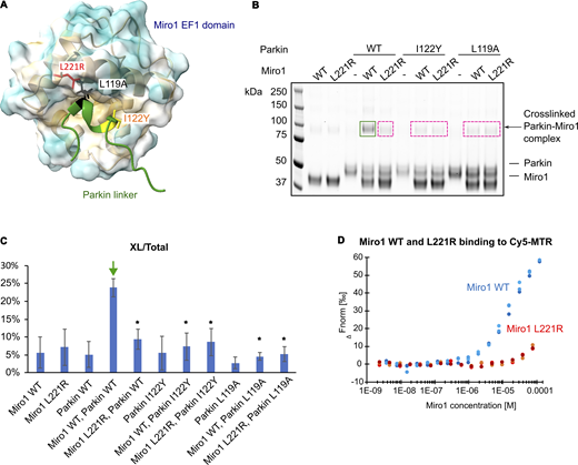
Substitutions in the MTR and Miro1 disrupt Miro1 binding to Parkin. (A) Positions of the mutated residues in the EF1 domain of Miro1 (surface) (L221R, red) and in Parkin linker (green ribbon) (I122Y, yellow, L119A, black) are indicated. (B) Cross-linking assay with mutated Miro1 and/or Parkin shows decrease in Miro1–Parkin complex formation (dashed pink boxes) as compared with WT Parkin and Miro1 (green box). (C) Quantification of cross-linked species as percentage of the total protein signal from B. Green arrow indicates the control experiment; asterisks indicate conditions with Parkin and Miro1 for which the P value was below 0.05 (from two-sided t test of at least three replicates). Error bars represent standard deviations from at least three experiments. (D) Binding of fluorescent synthetic peptide from Parkin linker region containing MTR (Cy5-MTR) to Miro1 WT (blue) and L221R mutant (red) was assayed using MST. Dots represent technical replicates. Affinity of Miro1 L221R mutant to Cy5-MTR was decreased, although affinities could not be calculated. Source data are available for this figure: SourceData F7.
Substitutions in the MTR and Miro1 disrupt Miro1 binding to Parkin. (A) Positions of the mutated residues in the EF1 domain of Miro1 (surface) (L221R, red) and in Parkin linker (green ribbon) (I122Y, yellow, L119A, black) are indicated. (B) Cross-linking assay with mutated Miro1 and/or Parkin shows decrease in Miro1–Parkin complex formation (dashed pink boxes) as compared with WT Parkin and Miro1 (green box). (C) Quantification of cross-linked species as percentage of the total protein signal from B. Green arrow indicates the control experiment; asterisks indicate conditions with Parkin and Miro1 for which the P value was below 0.05 (from two-sided t test of at least three replicates). Error bars represent standard deviations from at least three experiments. (D) Binding of fluorescent synthetic peptide from Parkin linker region containing MTR (Cy5-MTR) to Miro1 WT (blue) and L221R mutant (red) was assayed using MST. Dots represent technical replicates. Affinity of Miro1 L221R mutant to Cy5-MTR was decreased, although affinities could not be calculated. Source data are available for this figure: SourceData F7.
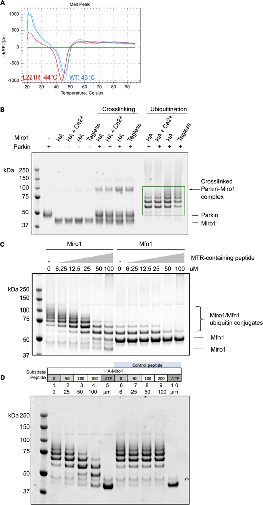
Effects of Miro1 L221R substitution, presence of HA tag, calcium binding, and MTR-containing peptide. (A) Differential scanning fluorimetry was performed on WT Miro1 and Miro1_L221R to assess the impact of the substitution on the thermal stability of the protein. Melting temperatures are indicated. (B) Cross-linking and ubiquitination assay was performed on WT Miro1 as compared with WT Miro1 preincubated with CaCl2 or to a tagless Miro1 with no HA tag. HA: Miro1 construct with an HA tag at the N terminus; tagless: Miro1 construct with no tag; Ca2+: Miro1 preincubated with CaCl2. Arrow indicates no change in detected cross-linked Parkin–Miro1 complex. Green box indicates no difference in ubiquitination of Miro1 with or without the HA tag or preincubated with CaCl2. (C) Excess of a synthetic MTR-containing peptide decreases Miro1 ubiquitination by Parkin. Miro1 or Mfn1 ubiquitination assays were performed with increasing concentrations of the MTR-containing peptide. (D) Ubiquitination assays with Miro1 and a control synthetic peptide. Source data are available for this figure: SourceData FS10.
Effects of Miro1 L221R substitution, presence of HA tag, calcium binding, and MTR-containing peptide. (A) Differential scanning fluorimetry was performed on WT Miro1 and Miro1_L221R to assess the impact of the substitution on the thermal stability of the protein. Melting temperatures are indicated. (B) Cross-linking and ubiquitination assay was performed on WT Miro1 as compared with WT Miro1 preincubated with CaCl2 or to a tagless Miro1 with no HA tag. HA: Miro1 construct with an HA tag at the N terminus; tagless: Miro1 construct with no tag; Ca2+: Miro1 preincubated with CaCl2. Arrow indicates no change in detected cross-linked Parkin–Miro1 complex. Green box indicates no difference in ubiquitination of Miro1 with or without the HA tag or preincubated with CaCl2. (C) Excess of a synthetic MTR-containing peptide decreases Miro1 ubiquitination by Parkin. Miro1 or Mfn1 ubiquitination assays were performed with increasing concentrations of the MTR-containing peptide. (D) Ubiquitination assays with Miro1 and a control synthetic peptide. Source data are available for this figure: SourceData FS10.
We first assayed Parkin variants L119A, I122Y, and Miro1 L221R in our cross-linking assay. Strikingly, these single amino acid substitutions substantially decrease formation of the cross-linked complex (Fig. 7, B and C). Next, we tested the binding of a fluorescently labeled MTR-containing peptide (Cy5-MTR) to Miro1 by microscale thermophoresis (MST). While we are unable to reach binding saturation and conclude binding affinity due to the limitations of the method for studying low-affinity interactions, we can nonetheless observe differences in the binding profile of WT versus L221R Miro1, with the substituted Miro1 binding compromised (Fig. 7 D).
Next, we wanted to verify whether Parkin ubiquitination of Miro1 is indeed dependent on the MTR interaction with the hydrophobic pocket in the Miro1 EF1 domain. We therefore set up a ubiquitination assay with mutated Parkin and Miro1. Substitutions L119A and I122Y in Parkin indeed cause a significant decrease in Miro1 ubiquitination (Fig. 8 A), suggesting that a single amino acid change in the MTR is sufficient to alter Parkin’s ability to ubiquitinate Miro1. While Miro1 is still detectably ubiquitinated by Parkin variants, the extent of modification is limited, suggesting that the disruption in Parkin–Miro1 binding impacts on the number of ubiquitins that Parkin conjugates to Miro1. Importantly, the Parkin L119A and I122Y protein do not exhibit any change in the levels of ubiquitination of a control substrate, Mfn1, indicating that these substitutions affect Miro1 ubiquitination specifically but do not influence the Parkin activity toward substrates in general. As for Miro1, the L221R variant is not ubiquitinated as efficiently as the WT and the reaction is slower (Fig. 8, A–C). Importantly, when a Parkin variant and Miro1 L221R variant were tested in the same reaction, the decrease of Parkin activity toward Miro1 was not significantly more pronounced as compared with the reaction with either Parkin or Miro1 mutant (Fig. 8 A). The fact that the effects of the mutations are not additive indicates the mutagenesis affects the same interaction site, providing further support for the interaction between MTR and the hydrophobic EF1 pocket in Miro1.
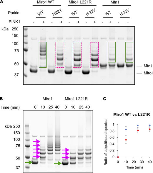
Substitutions in the MTR and Miro1 disrupt Miro1 ubiquitination by Parkin. (A) Miro1 WT and L221R mutant or Mfn1 (control) as indicated were subjected to ubiquitination assay with Parkin WT and/or variant I122Y or L119A. Decreased Miro1 ubiquitination as compared with WT (green) was marked with dashed pink boxes. (B) Time-resolved comparison of ubiquitination of Miro1 WT and L221R. Green arrows indicate unmodified Miro1, and pink arrows indicate ubiquitinated species of Miro1. (C) Quantification of ubiquitinated species of Miro1 WT and L221R over time based on at least three replicate experiments reveals slower kinetics of Miro1 L221R ubiquitination as compared with the WT Miro1. Error bars represent standard deviations from three experiments. Source data are available for this figure: SourceData F8.
Substitutions in the MTR and Miro1 disrupt Miro1 ubiquitination by Parkin. (A) Miro1 WT and L221R mutant or Mfn1 (control) as indicated were subjected to ubiquitination assay with Parkin WT and/or variant I122Y or L119A. Decreased Miro1 ubiquitination as compared with WT (green) was marked with dashed pink boxes. (B) Time-resolved comparison of ubiquitination of Miro1 WT and L221R. Green arrows indicate unmodified Miro1, and pink arrows indicate ubiquitinated species of Miro1. (C) Quantification of ubiquitinated species of Miro1 WT and L221R over time based on at least three replicate experiments reveals slower kinetics of Miro1 L221R ubiquitination as compared with the WT Miro1. Error bars represent standard deviations from three experiments. Source data are available for this figure: SourceData F8.
Synthetic MTR-containing peptides compete for Miro1 binding and decrease Miro1 ubiquitination
We reasoned that if the MTR is the main site of Parkin interaction with Miro1, the addition of a synthetic peptide that contains the MTR sequence should compete for the Parkin-binding site on Miro1. To test this, we synthesized an MTR-containing peptide (Parkin residues D115–R128) and added it to our cross-linking and activity assays. As expected, we observe a decrease in Parkin–Miro1 binding upon adding excess MTR peptide, as detected by cross-linking (Fig. 9, A and B). We also observe a decrease in Miro1 ubiquitination by Parkin in an activity assay (Fig. 9 C, peptide 1), further supporting the hypothesis that the excess of synthetic MTR peptide interferes with Parkin–Miro1 binding and subsequent Miro1 ubiquitination. At high concentrations of peptide, we also observe a slight decrease in Parkin activity toward Mfn1, which suggests MTR may have some additional role in Parkin activity regulation or participate in targeting of Mfn1, although to a much lower extent than in targeting Miro1 (Fig. S10 C). To verify that the inhibition is not due to chemical contamination from the peptide synthesis, we tested another unrelated peptide acquired from the same source. We do not observe any effect on Miro1 ubiquitination with this control peptide (Fig. S10 D).
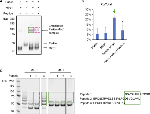
Competition with synthetic peptides containing MTR decreases Miro1 binding and ubiquitination by Parkin. (A) Cross-linking assay with Parkin and Miro1 in the presence of the MTR-containing peptide shows decrease of Parkin–Miro1 complex formation, indicated by the pink dashed box. Green box indicates the cross-linked band in a control experiment with no additional peptide. (B) Quantification of cross-linked species as percentage of the total protein signal from A. Error bars represent standard deviations from at least three experiments. Green arrow indicates the control experiment; asterisks indicate conditions with Parkin and Miro1 for which the P value was below 0.05 (from two-sided t test of at least three replicates). (C) Miro1 ubiquitination assay with a series of synthetic MTR-containing peptides. Dashed pink boxes indicate decreased Miro1 ubiquitination, as compared with control and unaffected reactions marked with green boxes. The sequences of corresponding peptides are indicated on the right. Ubiquitination assay with Mfn1 was used for comparison of Parkin activity in the presence of peptides. Green box indicates the residue stretch DSVGLAVIL in the MTR-containing peptide, responsible for decreased Miro1 ubiquitination by Parkin. Source data are available for this figure: SourceData F9.
Competition with synthetic peptides containing MTR decreases Miro1 binding and ubiquitination by Parkin. (A) Cross-linking assay with Parkin and Miro1 in the presence of the MTR-containing peptide shows decrease of Parkin–Miro1 complex formation, indicated by the pink dashed box. Green box indicates the cross-linked band in a control experiment with no additional peptide. (B) Quantification of cross-linked species as percentage of the total protein signal from A. Error bars represent standard deviations from at least three experiments. Green arrow indicates the control experiment; asterisks indicate conditions with Parkin and Miro1 for which the P value was below 0.05 (from two-sided t test of at least three replicates). (C) Miro1 ubiquitination assay with a series of synthetic MTR-containing peptides. Dashed pink boxes indicate decreased Miro1 ubiquitination, as compared with control and unaffected reactions marked with green boxes. The sequences of corresponding peptides are indicated on the right. Ubiquitination assay with Mfn1 was used for comparison of Parkin activity in the presence of peptides. Green box indicates the residue stretch DSVGLAVIL in the MTR-containing peptide, responsible for decreased Miro1 ubiquitination by Parkin. Source data are available for this figure: SourceData F9.
To further dissect the region in the MTR peptide sequence that is important for Miro1 binding and its subsequent ubiquitination, we tested a series of MTR-containing peptides covering Parkin residues E98–R128 (also containing the ACT element and the GLAVIL motif [Gladkova et al., 2018]) in Miro1 activity assays (Fig. 9 C). Only peptides that contain residues D115–L123 (DSVGLAVIL) are inhibitory, and the presence of the ACT element residues before (E98–G114) does not have any detectable effect.
Conservation of the Miro1 hydrophobic pocket and evolution of Parkin linker region
Having established the binding site between Parkin and Miro1, we wanted to further investigate the residues that are important for the interaction. For this, we looked at conservation of the Miro1 hydrophobic pocket. Human cells have two Miro isoforms—Miro1 and Miro2, which only differ by two residues in their Parkin-binding pocket, with no obvious change in the global shape or hydrophobicity of the pocket (Fig. S11 A). Consequently, AlphaFold still predicts confident binding of MTR to Miro1 EF1 domain (Fig. S11 D). Drosophila melanogaster (Dm) cells possess only one Miro isoform with a fold and distribution of hydrophobic residues in the Parkin-binding pocket very similar to human Miro1/2 (Klosowiak et al., 2013) (Fig. S11 B). However, DmParkin is not predicted to bind to DmMiro because MTR is not conserved in DmParkin (Figs. 6 C and S11 D). Interestingly, AlphaFold does predict DmMiro to bind to human Parkin, as it does even for the yeast Miro homolog, Gem1p (Frederick et al., 2004) (Fig. S11 D). In addition, many other Miro1 interactors have been predicted to bind to the same pocket besides Parkin (Covill-Cooke et al., 2024). Together, these data suggest that the interactive, “promiscuous” pocket in Miro EF1 domain is evolutionarily conserved, while Parkin linker evolved more recently (Fig. 6 C).

Structural overlay and protein sequence alignment of the EF1 domain of Miro protein homologs. (A) Overlay of human (Hs) Miro1 (purple) and Miro2 (red) EF1 domains. Residues from the Parkin-binding pocket that are different in Miro1 versus Miro2 are indicated in orange. (B) Overlay of human Miro1 (purple) and DmMiro (black) EF1 domains. Residues from the Parkin-binding pocket that are different in HsMiro1 versus DmMiro are indicated in orange. (C) Protein sequence alignments of the EF1 domains of Miro proteins (HsMiro1/2 and DmMiro). Top panel illustrates hydrophobicity of residues, and bottom panel highlights conserved residues. Orange boxes show differences in residues between the Miro1 proteins in the Parkin-binding pocket. (D) PAE plots from AlphaFold predictions for interaction of human Parkin (HsParkin) with HsMiro2, DmMiro, and yeast ScGEM1 show no confidence in predicted models (no interaction). (E) PAE plots from AlphaFold predictions for interaction of HsParkin with EF-hand domain-containing proteins (calmodulin 1, centrin 2, and calcineurin subunit B type 1) show no confidence in predicted models (no interaction). PAE, predicted aligned error.
Structural overlay and protein sequence alignment of the EF1 domain of Miro protein homologs. (A) Overlay of human (Hs) Miro1 (purple) and Miro2 (red) EF1 domains. Residues from the Parkin-binding pocket that are different in Miro1 versus Miro2 are indicated in orange. (B) Overlay of human Miro1 (purple) and DmMiro (black) EF1 domains. Residues from the Parkin-binding pocket that are different in HsMiro1 versus DmMiro are indicated in orange. (C) Protein sequence alignments of the EF1 domains of Miro proteins (HsMiro1/2 and DmMiro). Top panel illustrates hydrophobicity of residues, and bottom panel highlights conserved residues. Orange boxes show differences in residues between the Miro1 proteins in the Parkin-binding pocket. (D) PAE plots from AlphaFold predictions for interaction of human Parkin (HsParkin) with HsMiro2, DmMiro, and yeast ScGEM1 show no confidence in predicted models (no interaction). (E) PAE plots from AlphaFold predictions for interaction of HsParkin with EF-hand domain-containing proteins (calmodulin 1, centrin 2, and calcineurin subunit B type 1) show no confidence in predicted models (no interaction). PAE, predicted aligned error.
On the other side, we asked whether MTR in Parkin could interact with proteins that possess similar EF-hand fold as Miro1/2. There are many other EF-hand domain-containing proteins, and FoldSeek’s search (van Kempen et al., 2024) for human proteins that have similar folds to Miro1/2 EF1 domain yielded several proteins, including calmodulin, centrin, and calcineurin. Similarly to Miro1, these are calcium-regulated proteins with EF-hand domains. Using AlphaFold, we checked whether Parkin can be predicted to bind to these proteins, but no confident interaction is predicted (Fig. S11 E). Therefore, the MTR in Parkin seems to have evolved to interact specifically with Miro1/2 but not to other EF domains.
Discussion
Several studies have reported a surprisingly large number of proteins identified as ubiquitinated by Parkin under mitophagy-inducing conditions (Antico et al., 2021; Ordureau et al., 2014; Sarraf et al., 2013). However, it has remained unknown whether all identified proteins are direct Parkin substrates, and if yes, how Parkin recognizes and ubiquitinates such a diverse range of proteins. Moreover, no substrate recognition motif has been found in Parkin. Here, we show that full-length, untagged Parkin activated by PINK1 acts in a promiscuous manner and can directly ubiquitinate many different proteins in a biochemical assay. We establish that Miro1 is the most efficiently ubiquitinated substrate from our selected proteins in vitro, including ubiquitin and Parkin itself, suggesting that Parkin does have some level of specificity, at least in an in vitro reconstituted assay. It remains to be determined whether Parkin modifies Miro1 to a similar extent and with similar kinetics in a cellular context and how Parkin ubiquitinates other proteins besides the ones tested in this work. Previous reports did not agree on which substrates are preferentially ubiquitinated by Parkin under mitochondrial depolarization, but suggested mitofusins and voltage-dependent anion-selective channel proteins as candidates (Ordureau et al., 2018; Vranas et al., 2022). On the other hand, Mfn1 and Miro1 levels were found to be decreased in Parkin- and p97-dependent degradation upon mitochondrial depolarization, potentially suggesting that Mfn1 and Miro1 could be preferentially targeted for ubiquitination by Parkin (Ordureau et al., 2020). In our study, we used a truncated version of Mfn1 and it is possible that a full-length protein may be more favorably ubiquitinated by Parkin in the cell.
Further, despite the challenges encountered due to the low affinity of the interaction, through a series of biochemical approaches, we demonstrate the existence of an elusive Parkin–substrate interaction. We show that Parkin binds to Miro1 via a short hydrophobic region in the mostly disordered linker between Ubl and the RING0 domain of Parkin, in the same region that was recently suggested through AlphaFold modelling (Covill-Cooke et al., 2024). In addition, the same study shows that Parkin recruitment to mitochondria upon Miro1 overexpression was diminished upon L119A substitution, supporting the evidence for this linker region to be involved in Parkin-Miro1 interaction also in the cellular context. Our findings highlight the functional importance (in biochemical terms) of this moderately conserved region of Parkin, so far “invisible” in structures determined using x-ray crystallography due to its disordered nature, with the potential for conditional folding predicted computationally. Knowing that Parkin can detectably interact with Miro1, it is now tempting to speculate that Parkin could interact with other, not yet investigated proteins via the same or a different interaction site.
Since we used a truncated form of Miro1 that does not contain the N-terminal GTPase domain or the C-terminal transmembrane domain, we can only speculate about a possible involvement of these elements in Parkin binding. AlphaFold does not predict any contribution of the N-terminal GTPase domain in the Parkin–Miro1 interaction, positions the N-terminal GTPase domain flexibly connected to the rest of Miro1, and the only confidently predicted interaction region remains the same as in our Miro1 construct (Fig. S9, D and E). Of note, Miro1 overexpression and silencing experiments that indicate the existence of Parkin–Miro1 interaction in cells use full-length Miro1, suggesting the N-terminal GTPase domain does not disrupt Parkin interaction with Miro1 (Safiulina et al., 2018). Interestingly, the residue S156 in the N-terminal GTPase domain of Miro1 has been reported to be phosphorylated by PINK1 and to enhance Parkin translocation to the mitochondrial membrane (Shlevkov et al., 2016), suggesting that the modifications in the N-terminal GTPase domain in Miro1 might play a role in regulating Miro1 binding to Parkin.
The Parkin preference for targeting Miro1 as compared with its close homolog Miro2 was previously described in detail and attributed to the specific chemical environment of the favored lysine in the cGTPase domain of Miro1 (Klosowiak et al., 2016). While the cGTPase domain on its own is sufficient and necessary for Miro1 ubiquitination by Parkin, the presence of the EF1 domain leads to a slight increase in Miro1 and Miro2 ubiquitination, which we can now explain by increase in affinity resulting from Parkin binding to EF1. What emerges is a two-step Miro1 preference by Parkin: initially driven by the EF1-Parkin binding, and upon Parkin activation, by targeting a specific lysine on the cGTPase domain of Miro1.
Importantly, our results agree with cellular findings that Parkin can interact with Miro1 independently of its activation state (Safiulina et al., 2018; López-Doménech et al., 2021), as Parkin phosphorylation does not detectably increase its interaction with Miro1 based on cross-linking data. Together with the two-step preference for Miro1, this would suggest that Parkin binding to a substrate and its ubiquitination activity are uncoupled, and, at least in this specific case, Parkin activity is not induced by substrate binding. Parkin interaction with Miro1 has been previously observed in cellular studies, where Parkin translocation to mitochondria upon mitochondrial damage was dependent on Miro1/2 expression but occurred independently of PINK1 expression and, consequently, of Parkin activation state (Safiulina et al., 2018). This suggests that Miro1 could serve as a mitochondrial acceptor for Parkin even before activation of the PINK1/Parkin cascade, which generates pUb chains considered to be the main Parkin receptors (Okatsu et al., 2015; Vranas et al., 2022; Dunkerley et al., 2022; Koyano et al., 2019a), and does not get ubiquitinated until mitochondria are damaged and Parkin is activated. Consequently, a small pool of Parkin could be constantly present at the mitochondrial membrane, which has been indeed observed in cells (Narendra et al., 2008; Darios et al., 2003; Safiulina et al., 2018; Shlevkov et al., 2016). While the significance of this is unclear, we speculate that low levels of Parkin at the mitochondrial membrane, readily available upon PINK1-induced activation for a rapid ubiquitination of substrates and further Parkin recruitment through the feed-forward mechanism (Ordureau et al., 2014), could allow for a rapid response to mitochondrial damage. This may be important in dopaminergic neurons that are susceptible to mitochondrial stressors and where fast mitochondrial turnover is critical to prevent neurodegeneration (Haddad and Nakamura, 2015). While no pathogenic mutations in the Miro1-interacting site in Parkin have been reported so far, ClinVar database (Rehm et al., 2015) lists two missense mutations of uncertain significance (D115G and G118A), identified in patients with autosomal recessive juvenile PD, cancer, and inborn genetic diseases.
On the other side, the hydrophobic Miro1 pocket has been proposed to accommodate various other partners besides Parkin (Covill-Cooke et al., 2024), which suggests Parkin may be competing with other proteins, such as cytoskeletal adaptors CENPF, Trak, and MYO19, for binding to Miro1 in cells. A high local concentration of Parkin induced by the presence of pUb upon mitochondrial damage could lead to enhanced occupancy of Miro1 pocket by Parkin and result in disruption of Miro1 interactions with other binders. Ultimately, this could be the mechanism by which Miro1 is uncoupled from microtubules, leading to disconnected mitochondria with arrested motility that can be quarantined (Wang et al., 2011) or undergo mitophagy.
Our work brings a new perspective on how Parkin recognizes one of its substrates, and how it may be involved in mitochondrial homeostasis. This study may influence directions for therapeutic avenues in neurological disorders where mitochondrial dynamics are affected, such as in PD.
Materials and methods
Protein expression and purification
His6-Mfn1 (1–364-GSGSGSGGS-694–741), HA-Miro1 (181–579), His6-mitoNEET (33–108), FancI/D2, GST-PINK1 (126-C), Ube2L3, pUb, and Ub-MES were purified as previously described (Kumar et al., 2015; Conlan et al., 2009; Yan et al., 2018; El Oualid et al., 2010; Chaugule et al., 2019). In brief, His6-Mfn1, His6-mitoNEET, Ube2L3, and Ub constructs were expressed in BL21(DE3) Escherichia coli cells and purified using NiNTA affinity chromatography with His Pur Ni-NTA resin (cat. n 88222; Thermo Fisher Scientific) and Superdex 75 10/300 SEC. Ub was phosphorylated using GST-PINK1 construct. GST-PINK1 construct was purified using GST affinity chromatography with Pierce Glutathione Agarose (cat. n 16101; Thermo Fisher Scientific). Ub-MES was generated in a reaction with E1 enzyme and ATP at 37°C for 6 h prior to Superdex 75 10/300 SEC. FancI and FancD2 were expressed using Sf21 cells and purified using NiNTA affinity chromatography, ion exchange (HiTrap Q HP), and Superose 6 Increase 10/300 GL SEC.
Parkin was produced using modified protocols (Chaugule et al., 2011). Briefly, codon-optimized His6-Smt3-Parkin constructs were expressed in E. coli BL21(DE3) Rosetta cells at 37°C to an OD600 of 0.7 in 2xYT media supplemented with 0.5 mM ZnCl2. Expression was induced at 16°C with 300 μM IPTG for 18 h. After Ni2+NTA affinity purification, the His6-Smt3 tag was cleaved with Ulp1 protease overnight at 4°C, followed by ResourceQ ion exchange chromatography and Superdex 75 10/300 SEC. For pParkin, after the first purification step with Ni2+NTA affinity, Parkin was eluted and phosphorylated with PhPINK126-C while dialyzing in phosphorylation buffer (5 mM ATP, 5 mM MgCl2, 50 mM NaCl, 50 mM Tris-HCl, pH 8, and 1 mM DTT) for 1 h at 20°C. pParkin was then rebound to Ni2+NTA resin and further purified as for non-phosphorylated Parkin.
HA-Miro1181–579 was purified as follows: E. coli BL21 (DE3) cells were grown in 2xYT media until OD600 0.7, cooled down to 18°C, and induced with 300 μM IPTG for 18 h. Cells were lysed by sonication in lysis buffer (PBS with additional 200 mM NaCl, 0.4 mM TCEP, and 5% glycerol) supplemented with 4 mM MgCl2, protease inhibitor tablets (Pierce; Thermo Fisher Scientific), and DNAse I (Merck). After lysate clearing by centrifugation, GST-tagged Miro1 construct was purified on GSH agarose, and the tag was cleaved using 3C protease overnight at 4°C. Miro1 was then concentrated and purified on Superdex 75 10/300 SEC column.
Amino acid substitutions in Parkin and Miro1 were generated by site-directed mutagenesis, confirmed by sequencing, and purified as above. The following primers were used: for Miro1 L221R: forward 5′-CAACACTCCACGCGCTCCTCAAGC-3′, reverse 5′-AAACAAATCCTCTGAAAGAAG-3′; for Parkin L119A: forward 5′-TAGCGTTGGTGCGGCAGTTATTC-3′, reverse: 5′-TCACCAGGCAGAACG-3′; and for Parkin I122Y: forward 5′-TAGCGTTGGTTATGCAGTTATTCTGCATAC-3′, reverse: 5′-TCACCAGGCAGAACGCTG-3′.
Commercial reagents
MTR-containing peptides were synthesized by Genosphere Biotechnologies with >95% purity as assessed by HPLC and mass spectrometry and dissolved in 100% DMSO. Ubiquitin was purchased from Biotechne (U-100H) as lyophilized powder and dissolved in 100 mM NaCl and 20 mM HEPES, pH 7.4. DSS cross-linker was acquired from Thermo Fisher Scientific and dissolved in 100% DMSO.
Ubiquitination assays
For substrate ubiquitination assays, each substrate at 2 µM (besides FANCD2, which was at 1 µM due to a very strong signal due to high molecular mass) was mixed with 20 µM Ub, 0.1 µM E1, 0.5 µM Ube2L3, and 0.5 µM pParkin in ubiquitination buffer (50 mM Tris-HCl, pH 7.5, 100 mM NaCl, 5% glycerol, and 5 mM MgCl2). For analysis of ubiquitin consumption, assays were performed with 5 µM of each substrate, 0.2 µM pParkin, and 0.25 µM E1. Reactions were supplemented with 5 mM ATP where indicated and incubated for 40 min or as indicated at 37°C.
For Miro1 ubiquitination reactions with mutated Parkin and Miro1, 4 µM of Miro1 (or Mfn1 as control) was incubated with 20 µM Ub, 0.1 µM E1, 0.5 µM Ube2L3, 0.5 µM Parkin or pParkin as indicated, and 50 nM PINK1 where indicated, in ubiquitination buffer and ATP as above. Reactions were stopped after 45 min at 37°C.
For Miro1/Mfn1 reactions with synthetic peptides, 4 µM of Miro1 or Mfn1 was incubated with 20 µM of the reactive ubiquitin Ub-MES (to eliminate potential interactions of the peptide with E1 or E2) and 0.5 µM pParkin in ubiquitination buffer and ATP as above. 100 µM of peptide was added, unless specified otherwise, to a final of 2% DMSO, and the reaction was incubated for 1 h at 37°C.
The reactions were stopped by adding NuPAGE 4x LDS loading buffer (Novex) and 167 µM DTT prior to being resolved on NuPAGE LDS 4–12% Bis-Tris gels (Novex). Gels were stained with a Coomassie-based SimplyBlue SafeStain (Invitrogen) and scanned using the LI-COR Odyssey Clx Infrared Imaging System at a 700 nm wavelength. For western blotting, the proteins resolved by SDS-PAGE were transferred onto nitrocellulose membranes using an iBlot gel transfer device (Invitrogen), blocked with 5% milk PBS-T (0.1% Tween 20), and incubated with primary antibodies overnight at 4°C. The following antibodies were used: anti-HA (51064-2-AP; ProteinTech), anti-Parkin (1A1; IBL; 14060-1-AP; ProteinTech), anti-Mfn1 (13798-1-AP; ProteinTech), anti-His6 (66005-1-Ig; ProteinTech), and anti-FANCD2 (33539; DU) at 1:1,000 dilutions. The following day, the membranes were washed with PBS-T and revealed using secondary LI-COR antibodies at 1:20,000 dilutions: IRDye 680RD Donkey anti-Rabbit IgG (cat. n 926–68073), IRDye 800CW Donkey anti-Mouse IgG (cat. n 926–32212), and IRDye 800CW Donkey Anti-Goat IgG (cat. n 926–68074). The membranes were then washed with PBS-T before scanning using the LI-COR Odyssey Clx Infrared Imaging System at 700- and 800 nm wavelengths. For quantification, the band densitometry was measured from at least three replicates, and a t test was applied to calculate P values.
Cross-linking and mass spectrometry
3 µM of Parkin constructs were mixed with 3 µM HA-Miro1181–579 in 50 mM HEPES, pH 7.4, 150 mM NaCl, 0.4 mM TCEP, and 5% glycerol in a 10 μl volume and preincubated for 5 min at room temperature (20°C). Then 0.3 mM of DSS cross-linker (Thermo Fisher Scientific) in DMSO was added, and the reaction was incubated for a further 45 min. Reaction was stopped by adding 50 mM Tris-HCl, pH 8. The samples were resolved on a NuPAGE LDS 4–12% Bis-Tris gels (Novex) under reducing conditions and Coomassie stained. Bands corresponding to expected Parkin–Miro1 complex size (∼100 kDa) were excised and sent for mass spectrometry (BSRC Mass Spectrometry & Proteomics Facility, University of St. Andrews, St. Andrews, UK). The samples were reduced, alkylated, and trypsin-digested at the facility, and the resulting peptides were analyzed by liquid chromatography-tandem mass spectrometry (LC-MSMS) on an Orbitrap Fusion Lumos mass spectrometer (Thermo Fisher Scientific) using collision induced dissociation and electron transfer dissociation (CID+ETD) activation. The cross-linked peptides were identified using MeroX 2.0.1.4 software (Götze et al., 2012; Götze et al., 2015) with default settings (1% false discovery rate, 10% prescore intensity, and three missed cleavages allowed) with the following changes: Phosphorylation included as posttranslational modification, BS3/DSS cross-linker, and specificity site: lysines only.
SEC and SEC-SAXS
For the large-scale cross-linking and purification of the Parkin–Miro1 complex, the cross-linking reaction was performed as described above but in 10 ml final volume, in 50 mM HEPES, pH 7.4, 150 mM NaCl, with 0.2 mM DSS. After incubation and quenching, the sample was concentrated and purified on a Superdex 200 Increase 10/300 size exclusion column at 0.25 ml/min. Fractions from the peak corresponding to the Parkin-Miro1 complex were collected, analyzed by SDS-PAGE and western blotting, concentrated and flash-frozen in liquid nitrogen.
The samples were shipped to Diamond Light Source, beamline B21. Samples were applied onto a Superdex 200 Increase 3.2 column equilibrated in a buffer comprising 150 mM NaCl, 50 mM HEPES pH 7.4, 0.4 mM TCEP, and 5% glycerol at 0.16 ml/min before exposure to x-rays as part of the standard setup at the beamline. Data were analyzed using ScÅtter version IV.j (available from beamline B21 at Diamond Light Source). Ab initio models were calculated by 21 independent runs of DAMMIF (Franke and Svergun, 2009), followed by averaging and filtering using DAMAVER (Volkov and Svergun, 2003).
AlphaFold predictions using ColabFold and model visualizations
ColabFold (Mirdita et al., 2022; Jumper et al., 2021) version 1.5.2.20231005 has been used through the MVLS Advanced Research System computing platform at the University of Glasgow with the following parameters: Number of recycles: 12; number of models: 5, with amber relaxation. Models were visualized using PyMOL (The PyMOL Molecular Graphics System, Version 3.0 Schrödinger; LLC) or with UCSF ChimeraX, developed by the Resource for Biocomputing, Visualization, and Informatics at the University of California, San Francisco, with support from National Institutes of Health R01-GM129325 and the Office of Cyber Infrastructure and Computational Biology, National Institute of Allergy and Infectious Diseases (Goddard et al., 2018).
NMR experiments
The autoinhibitory Ubl domain and linker region of human Parkin (residues 1–126, Ubl1–126) was created by inserting a stop codon in full-length Parkin. Ubl1–126 was expressed as a His6-SUMO fusion protein in E. coli BL21(DE3) cells using M9 minimal media supplemented with 15NH4Cl (1.0 g/liter) for NMR experiments and purified as previously reported (Aguirre et al., 2017; Chaugule et al., 2011). Samples of 15N-labeled Ubl1–126 (140 µM) were prepared in 50 mM Tris, 150 mM NaCl, and 250 µM TCEP (pH 7.5) with 10% (vol/vol) D2O. Imidazole was included as a pH indicator and 4,4- dimethyl-4-silapentane-1-sulfonic acid as a direct internal 1H reference. 1H-15N HSQC spectra were acquired at 25°C on a Bruker Avance Neo 600 MHz NMR spectrometer equipped with a triple resonance probe using z-field gradients (BioCORE Facility, Western University). Spectra were collected in the absence and presence of an equimolar amount of Miro1180–582. Data were processed using NMRPipe (Delaglio et al., 1995), visualized using NMRViewJ (Johnson and Blevins, 1994), and assigned using previously reported chemical shift data (Aguirre et al., 2017).
MST
For the MTR-containing peptide-binding experiments to Miro1, fluorescent Cy5-MTR peptide at the final concentration of 20 nM in PBS with 2.5% DMSO, 0.05% Tween 20, and 0.5 mg/ml bovine serum albumin was titrated with Miro1 WT or Miro1 L221R. Measurements were performed at 18–22°C on a Monolith NT.115 instrument (NanoTemper Technologies) using the red channel with 20% excitation power and 40% MST power. Data were analyzed using the MO Affinity Analysis v2.3 (NanoTemper Technologies).
Online supplemental material
Fig. S1 shows the additional assays with Parkin mitochondrial substrates; Fig. S2 shows the results from orthogonal methods to capture Parkin–Miro1 complex; Fig. S3 shows the additional information about the SAXS data of Parkin–Miro1 complex; Figs. S4, S5, S6, S7, and S8 show the additional mass spectrometry data; Fig. S9 shows the additional AlphaFold models of Parkin–Miro1 complex; Fig. S10 shows the control experiments with Miro1 L221R variant and MTR-peptide assays; and Fig. S11 shows the sequence alignments and AlphaFold predictions of EF1 domains from Miro1 and other proteins. Table S1 shows additional mass spectrometry data.
Data availability
Most data are available in the article itself and its supplementary materials. Additional data are available from the corresponding author upon reasonable request.
Acknowledgments
The authors would like to thank the BSRC Mass Spectrometry & Proteomics Facility, University of St. Andrews, St. Andrews, UK, for the mass spectrometry experiments; June Southall from the Neil Bulleid Integrated Protein Analysis Facility, Shared Research Facilities, University of Glasgow, for help with MST, isothermal titration calorimetry, and surface plasmon resonance experiments; and Diamond Light Source for beamtime for the SEC-SAXS experiments (BAG allocation mx28516). We also thank Connor Arkinson for making the UBE1 and FANCD2 proteins and Martin Rennie for helpful discussions.
This research was supported through grants from the Wellcome Trust (209347/Z/17/Z, H. Walden) and the Canadian Institutes of Health Research (PJT 166019, G.S. Shaw). For the purpose of open access, the authors have applied a Creative Commons Attribution (CC BY) license to any Author Accepted Manuscript version arising from this submission.
Author contributions: J. Koszela: conceptualization, data curation, formal analysis, investigation, methodology, validation, visualization, and writing—original draft, review, and editing. A. Rintala-Dempsey: investigation and writing—original draft, review, and editing. G. Salzano: conceptualization and investigation. V. Pimenta: formal analysis, investigation, methodology, validation, visualization, and writing—original draft. O. Kamarainen: investigation and writing—review and editing. M. Gabrielsen: formal analysis, investigation, and writing—review and editing. A.L. Parui: data curation, formal analysis, investigation, methodology, validation, and visualization. G.S. Shaw: conceptualization, funding acquisition, methodology, project administration, resources, supervision, validation, and writing—original draft, review, and editing. H. Walden: conceptualization, funding acquisition, methodology, supervision, and writing—review and editing.


