Centrioles are essential for centrosome and cilium formation. In fly spermatocytes, they undergo dramatic elongation to form giant centrioles that support sperm development. The conserved centriole protein Ana1/Cep295 is known to promote this elongation, yet the underlying mechanism remains poorly understood. Here, we show that Ana1 regulates centriole length through two distinct mechanisms. It directly interacts with Cep135 to promote centriole elongation and to recruit Cep135 into the proximal centriole-like structure. Ana1 also binds and bundles microtubules in vitro, and this activity is required for centriole elongation in vivo. Importantly, these two interactions are not mutually exclusive and both contribute to Ana1-induced centriole overelongation. Together, our findings reveal that Ana1 is involved in centriole elongation through interactions with distinct centriolar components during fly spermatogenesis.
Introduction
Centrioles are microtubule (MT)-based structures that are essential for the assembly of centrosomes and cilia/flagella (Azimzadeh, 2014). Centrosomes function as the primary MT-organizing centers in most animal cells and play pivotal roles in a variety of cellular processes, including cell division, cell differentiation, and the establishment of cell polarity (Bettencourt-Dias et al., 2011; Conduit et al., 2015; Nigg and Raff, 2009). In quiescent cells that enter the G0 phase, centrioles dock at the plasma membrane where they become basal bodies to initiate the formation of cilia and flagella (Dutcher, 2003). Defects in centriole assembly result in the malformation and dysfunction of these critical organelles, and have been linked to a diverse set of human pathologies. Therefore, elucidating mechanisms that govern centriole biogenesis and maintenance is crucial for understanding their function and the pathological consequences of their dysfunction.
Centriole assembly begins with the recruitment of the protein kinase ZYG1 (in worms) or Plk4 (in mammals and flies) to the outer wall of the mother centriole (Bettencourt-Dias et al., 2005; Habedanck et al., 2005; O’Connell et al., 2001; Sonnen et al., 2012), which triggers the hierarchical assembly of core cartwheel components including SAS5/STIL/Ana2 (worms, mammals, and flies, respectively) and Sas6 (Arquint et al., 2015; Kratz et al., 2015; Lettman et al., 2013; Moyer et al., 2015; Ohta et al., 2014). The resulting ninefold symmetric cartwheel structure then provides a scaffold for centriole formation (Banterle et al., 2021; Kitagawa et al., 2011; Laporte et al., 2024; van Breugel et al., 2011). Subsequent recruitment of Sas4/CPAP promotes the polymerization of centriolar MTs, which surround and stabilize the inner cartwheel structure (Cottee et al., 2013; Hatzopoulos et al., 2013; Hsu et al., 2008; Tang et al., 2009; Tang et al., 2011).
Following their assembly, centrioles recruit pericentriolar material (PCM) and mature into centrosomes. Recent studies have demonstrated that the recruitment of Cep295 (human)/Ana1 (Drosophila) is essential for centriole-to-centrosome conversion (Fu et al., 2016; Izquierdo et al., 2014; Tsuchiya et al., 2016). In human cell lines, depletion of Cep295 impairs the recruitment of PCM and leads to centriole destabilization (Izquierdo et al., 2014). In Drosophila, ana1 mutant flies are severely uncoordinated due to the lack of functional cilia and exhibit a significantly reduced number of centrosomes in third-instar larval brains (Blachon et al., 2009; Saurya et al., 2016). In cultured fly cell lines, Ana1 is recruited to centrioles during late anaphase and is required for the recruitment of Asl (Fu et al., 2016), which in turn promotes PCM recruitment and the subsequent maturation of centrioles into centrosomes. Asl also licenses the daughter centriole for duplication in the next cell cycle through the recruitment of the Plk4 kinase (Novak et al., 2014; Novak et al., 2016). In addition, Ana1 contributes to centrosome maintenance in oocytes, a function that depends on Polo kinase but is independent of the PCM (Pimenta-Marques et al., 2024).
Apart from its role in centriole-to-centrosome conversion, Ana1 has also been implicated in centriole elongation, particularly in the male germline where centrioles undergo dramatic growth during spermatogenesis (Alvarez-Rodrigo et al., 2021; Saurya et al., 2016). In fly spermatocytes, the centriole length is directly correlated with the expression level of Ana1: Ana1 overexpression induces the overelongation of centrioles, whereas reduced Ana1 dosage leads to the formation of shorter centrioles. However, the molecular mechanism underlying this function of Ana1 remains poorly understood. Here, we identified distinct regions within Ana1 that mediate interactions with Cep135 and MTs, and demonstrated that both activities are required for centriole length control. We further showed that the Ana1-Cep135 interaction is dispensable for the recruitment of Ana1 to centrioles, but is specifically required for loading Cep135 into the proximal centriole-like (PCL) structure during later stages of spermatogenesis, suggesting a context-dependent Ana1-Cep135 recruitment hierarchy.
Results
Ana1 is essential for centriole assembly and promotes centriole overelongation in fly spermatocytes
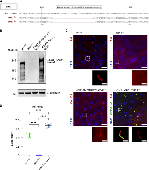
Previous studies of Ana1’s function in centriole and centrosome assembly have relied largely on the ana1mecB mutant. However, recent work has suggested that the truncated Ana1 protein encoded by this mutant allele may confound phenotypic interpretation through intragenic complementation (Nagy et al., 2025). To reexamine the functional role of Ana1 in fly spermatocytes, we therefore analyzed a CRISPR/Cas9-derived ana1 null mutant that lacks the entire coding region, including all four exons and three introns (Fig. 1, A and B; see Materials and methods for details). Unless otherwise stated, all experiments in this study were performed using this null mutant.
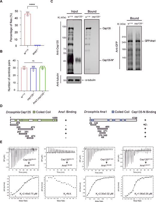
ana1 mutant flies were severely uncoordinated, likely due to defective centriole duplication and the consequent failure to form functional cilia in mechanosensory neurons (Fig. S1 A) (Blachon et al., 2009). In ana1−/− spermatocytes, Asl localization was completely abolished, consistent with its known dependency on Ana1 for centriolar recruitment (Fig. 1 C). Although Cep135-mRuby3–positive foci were occasionally observed, no morphologically recognizable centrioles could be detected (Fig. 1 C). These defects were fully rescued by the expression of EGFP-Ana1 under the control of a ubiquitin promoter (Fig. 1, B and C). Moreover, as previously reported, the overexpression of EGFP-Ana1 induced centriole overelongation in spermatocytes (Fig. 1, C and D). Together, our data confirmed that Ana1 is essential for centriole assembly in vivo and also established a robust genetic system to dissect mechanisms by which Ana1 promotes centriole elongation in fly spermatocytes.
Cep135 is required for Ana1-induced centriole overelongation
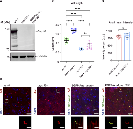
Previous studies have shown that loss of Cep135 leads to shorter centrioles and the absence of the central pair of MTs in flagellar axonemes (Blachon et al., 2009; Carvalho-Santos et al., 2012; Mottier-Pavie and Megraw, 2009; Roque et al., 2012). To determine whether Cep135 is required for Ana1-dependent centriole overelongation, we expressed EGFP-Ana1 in cep135c04199 mutant flies. Depletion of full-length Cep135, confirmed by western blotting (Fig. 2 A), strongly suppressed Ana1-induced centriole overelongation in primary spermatocytes, although these centrioles were marginally longer than those in cep135c04199 flies without Ana1 overexpression (Fig. 2, B and C). Consistent with previous reports (Blachon et al., 2009; Roque et al., 2012), centriole duplication appeared largely unaffected (Fig. S1 B). Notably, EGFP-Ana1 still localized robustly to centrioles in the absence of Cep135 (Fig. 2 B), and the amount of Ana1 recruited per unit length of centrioles was unchanged (Fig. 2 D). Since the cep135c04199 mutant is predicted to retain an N-terminal fragment (Fu et al., 2016; Mottier-Pavie and Megraw, 2009), we considered whether this residual protein might facilitate Ana1 recruitment. Pull-down experiments demonstrated that this fragment does not interact with Ana1 (Fig. S1 C). Taken together, these results indicate that Cep135 is required for Ana1-induced centriole overelongation but is dispensable for the initial recruitment of Ana1 to centrioles in fly spermatocytes.
Mapping of the Ana1-Cep135 interaction interface
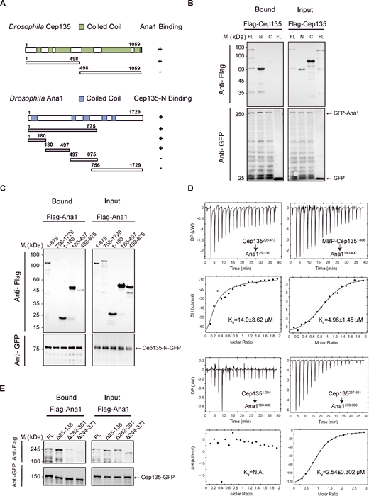
To determine whether Cep135 contributes to Ana1-induced centriole overelongation through a direct interaction, we set out to define the binding interface between the two proteins. Although Ana1 has previously been shown to interact with Cep135 in cultured D.Mel-2 cells (Fu et al., 2016), the underlying interface has remained poorly characterized. We therefore performed GFP pull-down experiments using a series of Ana1 and Cep135 fragments (Fig. 3 A). Consistent with previous findings, the full-length Ana1 protein showed robust binding to the N-terminal region of Cep135 (Fig. 3 B). Further mapping experiments revealed that Cep135 binds two distinct regions within Ana1, aa1–180 and 180–497 (Fig. 3 C).
We next measured the binding affinity between Cep135 and Ana1 by isothermal titration calorimetry (ITC)–based experiments using recombinantly purified MBP-tagged Cep1351–498 and the two fragments of Ana1 (aa25–138 and aa180–400) (Fig. 3 D). Cep1351–498 formed a 1:1 complex with Ana1180–400, with a dissociation constant (Kd) of ∼4.96 μm, whereas its interaction with Ana125–138 was weaker (Kd of ∼14.9 μm). Additional ITC-based measurements using shorter fragments of both proteins further identified Cep135257–351 and Ana1270–300 as the minimal binding regions, with a Kd of ∼2.54 μm (Fig. 3 D; and Fig. S1, D and E).
To evaluate the contribution of these two Ana1 regions to Cep135 binding, we made deletion constructs targeting each mapped interaction site and performed GFP pull-down assays. Deletion of the major Cep135-binding region (aa282–301/aa244–371) nearly abolished the interaction, whereas deletion of the weaker N-terminal region (aa25–138) had no detectable effect (Fig. 3 E). It should be noted that this weaker N-terminal region overlaps with the highly conserved CR1 domain (aa19–134), which is found in all CEP295/Ana1 homologs (Tsuchiya et al., 2016). Thus, although dispensable for Cep135 binding, its strong evolutionary conservation suggests that it may have other functional roles.
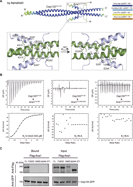
Having defined the major Cep135-binding region in Ana1, we next sought to gain structural insights into this interaction. We used AlphaFold3 (AF3)-based prediction (Abramson et al., 2024) to model the Cep135250–352-Ana1276–298 complex (Fig. 4 A). The five top-ranked solutions predicted highly similar models of a heterotetrameric complex, which is composed of an Ana1 dimer and a Cep135 dimer arranged in an antiparallel orientation (pTM = 0.66, ipTM = 0.65; Fig. S3 A). Evolutionarily conserved residues were enriched at the packing interface between the two proteins (Fig. 4 A and Fig. S2 A). In particular, the invariant residues Phe283, Ile286, and Val290 of Ana1 were buried within the hydrophobic core of the coiled-coil tetramer, while the invariant Arg282 and the highly conserved Glu294 residues were positioned to form electrostatic interactions with the adjacent Cep135 dimer.
To test the potential relevance of these predicted contacts, we substituted the two core hydrophobic residues in Ana1 with charged aa (F283D and I286D). Neither substitution appeared to drastically perturb the folding of Ana1 in vitro (Fig. S2 B), but both completely abolished Cep135 binding, as demonstrated by ITC-based experiments and GFP pull-down assays (Fig. 4, B and C). Together, these data strongly support the predicted contact interface between Ana1 and Cep135.
The Ana1-Cep135 interaction promotes centriole elongation
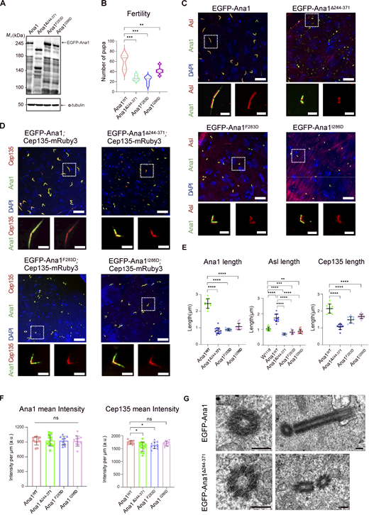
In order to investigate the functional importance of the Ana1-Cep135 interaction in vivo, we generated transgenic flies expressing either WT EGFP-Ana1 or Cep135 binding–deficient Ana1 mutants under the control of a ubiquitin promoter. These included a deletion mutant lacking the major Cep135-binding region (Ana1Δ244–371) and two single-point mutants, Ana1F283D and Ana1I286D, both of which abolished Cep135 binding in vitro. Western blotting confirmed that WT and mutant Ana1 proteins were expressed at similar levels (Fig. 5 A). Both EGFP-Ana1 and its binding-deficient mutants strongly rescued the severe uncoordination defect of ana1 mutant flies (Fig. S2 C), and all rescued flies were fertile. However, male flies that expressed the Cep135 binding–deficient Ana1 mutants produced significantly fewer pupae (Fig. 5 B). These results suggest that the Ana1-Cep135 interaction is dispensable for neurosensory cilium function but critical for germline function, particularly during spermatogenesis.
We next analyzed the behavior of WT and mutant EGFP-Ana1 proteins and their effects on centriole assembly in primary spermatocytes. All EGFP-Ana1 variants localized to centrioles marked by Asl, and centriole duplication appeared unaffected (Fig. 5 C and Fig. S2 D). The Cep135-mRuby3 signal was also detected along the entire centriole length in all genotypes (Fig. 5 D). Nevertheless, mutant centrioles were significantly shorter than WT controls and, notably, even shorter than those assembled from endogenous Ana1 (Fig. 5 E and Fig. S2 E). Thus, the Ana1-Cep135 interaction is required not only for Ana1-induced centriole overelongation but also for normal centriole length control. Quantification of Ana1 and Cep135 levels at centrioles revealed that Ana1 fluorescence intensity per unit length of centrioles was unchanged in all mutants, while Cep135 intensity was slightly reduced (Fig. 5 F). Transmission electron microscopy (TEM) analysis on preparations of WT and mutant testes further confirmed that despite their reduced lengths, no obvious structural defects were detected in the mutant centrioles analyzed (Fig. 5 G). Together, these findings demonstrate that the Ana1-Cep135 interaction is crucial for centriole overelongation and normal centriole length control, but is dispensable for the initial recruitment of Ana1 to centrioles.
The Ana1-Cep135 interaction promotes Cep135 incorporation into the PCL structure
Having established the role of the Ana1-Cep135 interaction in centriole elongation, we then asked whether this interaction also functions at a later stage of spermiogenesis. In late spermatids, centriole duplication factors are recruited proximal to the base of the centriole/basal body to form the PCL structure. The PCL structure contains core centriole proteins, including Plk4, Ana2, Sas6, Ana1, and Cep135, and serves as a template for daughter centriole formation in the early embryo (Blachon et al., 2009; Buglak et al., 2024). In both WT and mutant spermatids, PCL structures appeared synchronously within the spermatid cyst at the leaf nuclear stage, as marked by an EGFP-Ana1–positive bulge adjacent to one end of the centriole. RFP-Sas6 was robustly recruited to the PCL structure in all genotypes (Fig. 6, A and B). In contrast, Cep135-mRuby3 incorporation into the PCL structure was nearly abolished in spermatids that expressed binding-deficient mutants (Fig. 6, C and D). Thus, the Ana1-Cep135 interaction also contributes to the efficient recruitment of Cep135 into the PCL structure during spermatid maturation.
Ana1 binds and bundles MTs via its N- and C-terminal regions
Although the Ana1-Cep135 interaction is crucial for centriole overelongation, our earlier finding that Ana1 overexpression induced residual centriole elongation in cep135−/− spermatocytes (Fig. 2, B and C) raised the possibility that Ana1 may also act through additional mechanisms. Previously published expansion microscopy data have shown that Ana1 localizes to the centriolar wall (Tian et al., 2021), and we therefore speculated that Ana1 may directly associate with MTs.
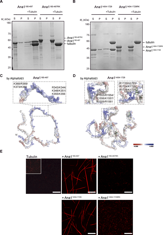
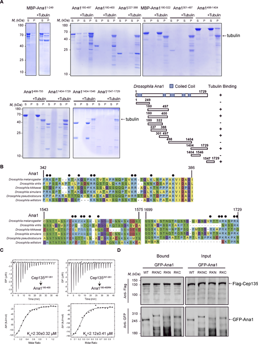
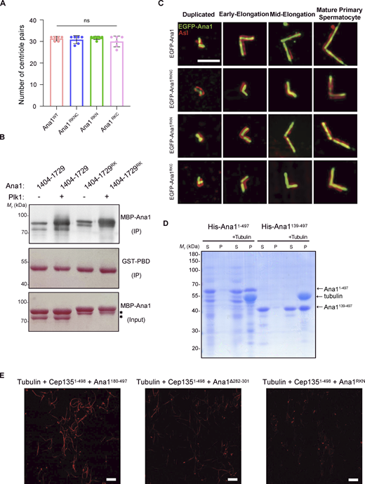
Since the full-length Ana1 protein could not be purified using a recombinant expression system, we instead purified Ana1 fragments and evaluated their ability to bind MTs using cosedimentation assays. We found two regions of Ana1, aa180–497 (hereafter Ana1 N fragment) and aa1404–1729 (hereafter Ana1 C fragment), efficiently coprecipitated with prepolymerized tubulin, indicating direct MT binding (Fig. 7, A and B; and Fig. S4 A). Fluorescence imaging further showed that both fragments could bundle MTs in vitro. In the presence of either fragment, the originally short and thin single MT filaments assembled into long, thick fascicles (Fig. 7 E).
To further characterize how Ana1 binds MTs, we analyzed AF3-predicted structural models of the N and C fragments. Surface charge analysis revealed one cluster of positively charged residues in the N fragment and two in the C fragment that are likely exposed for MT binding (Fig. 7, C and D; and Fig. S3 B). These charged patches are highly conserved among all Drosophila species (Fig. S4 B). To test their functional importance, we substituted these residues with Ala (referred to as the “RK” mutation). In both fragments, the RK mutations strongly impaired MT binding and bundling, as shown by cosedimentation and fluorescence imaging–based assays, respectively (Fig. 7, A, B, and E). Importantly, ITC-based experiments and GFP pulldowns both confirmed that these MT binding–deficient mutations had no influence on the Ana1-Cep135 interaction (Fig. S4, C and D).
The Ana1-MT interaction promotes centriole elongation in vivo
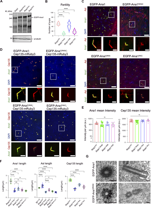
To test whether the MT-binding activity of Ana1 is essential for centriole elongation in vivo, we introduced RK mutations into EGFP-tagged full-length Ana1 protein to disrupt either the N-terminal site (hereafter referred to as Ana1RKN), the C-terminal site (hereafter referred to as Ana1RKC), or both (hereafter referred to as Ana1RKNC). All variants were expressed under a ubiquitin promoter and could fully rescue the uncoordination phenotype of ana1 mutant flies (Fig. 8 A and Fig. S2 C). While male fly fertility was partially restored in all genotypes, only Ana1RKN supported pupal production at near-WT levels (Fig. 8 B).
Using Asl and Cep135 as centriole markers, we found that MT binding–deficient Ana1 mutants strongly localized to centrioles and centriole duplication was not affected (Fig. 8 C and Fig. S5 B). However, these mutants failed to promote centriole overelongation to the same extent as WT Ana1 (Fig. 8, C and D; and Fig. S5 A). Further quantification revealed that the amounts of Ana1 and Cep135 recruited per unit length of centrioles were not significantly altered (Fig. 8 E), and therefore, the elongation defect was not likely due to impaired centriolar recruitment. The severity of centriole shortening followed a gradient: Ana1RKN showed the mildest effect, Ana1RKC a stronger reduction, and Ana1RKNC the most severe defect (Fig. 8 F). Furthermore, we compared the length of centrioles in spermatocytes expressing MT binding–deficient (Ana1RKNC) or Cep135 binding–deficient (Ana1Δ244–371) mutants, and found that the two types of mutations showed a comparable reduction in centriole length as measured by Asl (Fig. 8 F). And in either case, no obvious structural defects were detected, as confirmed by TEM (Fig. 8 G).
A recent study reported that Ana1 mutants deficient in Polo kinase binding also showed defects in centriole elongation (Alvarez-Rodrigo et al., 2021). To rule out the possibility that elongation defects observed in RK mutants were due to impaired Polo kinase interaction, we conducted GST pull-down experiments. It was demonstrated that the Ana11404–1729RK mutant protein retained full binding to the Polo-box domain (Fig. S5 C). We also examined whether the conserved CR1 region of Ana1, whose deletion had no detectable effect on Cep135 binding, might be involved in MT association. The CR1-deleted Ana1 protein could still cosediment with prepolymerized tubulin (Fig. S5 D).
Taken together, these results indicate that the MT-binding activity of Ana1 is required for centriole overelongation in vivo and that disruption of this activity leads to defects in elongation that are comparable in severity to those caused by disruption of the Ana1-Cep135 interaction.
Discussion
Ana1/CEP295 proteins are crucial for centriole-to-centrosome conversion and have been implicated in centriole elongation, but the mechanisms through which they drive giant centriole assembly during spermatogenesis remain poorly understood. Our study demonstrates that Ana1 promotes centriole elongation through two molecular activities—binding to Cep135 and direct association with MTs. Disruption of either interaction suppressed Ana1-induced centriole overelongation and reduced centriole lengths below the endogenous level. Intriguingly, both types of mutants converged on comparable centriole lengths, with ∼0.7227 μm for Ana1Δ244–371 centrioles and 0.6958 μm for Ana1RKNC centrioles as measured by the Asl signal. This observation led us to propose that there might be a shared structural limit imposed by the proximal centriolar scaffold, such as the cartwheel, the A-C linker, and/or the inner scaffold, which has been demonstrated to reinforce the MT wall (Cai et al., 2025; Guichard et al., 2013; Klena et al., 2020; Le Guennec et al., 2020; Li et al., 2019; Nazarov et al., 2020; Ruehle et al., 2024). Beyond this proximal core, continued longitudinal extension of the outer MT triplets likely requires additional scaffolding provided by the Cep135-Ana1 complex and by Ana1’s direct MT-binding activity. Flies expressing these binding-deficient Ana1 mutants exhibited no coordination defects, which suggests that neurosensory cilia remained functionally intact. Their male fertility was nonetheless only partially rescued, which underscores the importance of correct centriole lengths for gametogenesis.
We mapped MT binding to positively charged patches within the Ana11404–1729 region and showed that these residues are required for centriole elongation. In line with this, a recent study reported that Ana11472–1620 is required for centriole elongation, although the underlying mechanism was not elucidated (Nagy et al., 2025). Notably, deletion of this region completely abolished Ana1’s ability to rescue male fertility in ana1null flies, whereas our point mutants within the same region only partially impaired male fertility. This phenotypic difference suggests that the Ana11472–1620 region might mediate additional interactions beyond MT binding that also contribute to centriole function during spermatogenesis. In addition, Ana1 also binds MTs through its N-terminal region, although this contribution appears weaker. We speculate that these dual MT-binding interfaces may facilitate the longitudinal stacking of Ana1 and its associated complexes, thereby promoting the elongation of MT triplets during the formation of sperm-specific giant centrioles. In relation to this, human Cep295 binds centriolar MTs at the proximal end and stabilizes the MT wall independently of Cep135 (Atorino et al., 2020). It also promotes the assembly of the distal half of centrioles through recruitment of POC5 and POC1B, which then triggers posttranslational modification of centriolar MTs (Chang et al., 2016). Together, these findings suggest a conserved role of Ana1/CEP295 in direct MT binding and centriole elongation.
Cep135 has also been reported to bind MTs in vitro and in cultured cells (Carvalho-Santos et al., 2012). It is therefore plausible that Ana1, Cep135, and MTs stabilize one another within a ternary complex that reinforces the centriolar MT wall and supports continued longitudinal extension of the outer MT triplets. In support of this model, our in vitro reconstitution assay showed that a mixture of the Ana1 N fragment with MBP/Cep1351–498 promoted the formation of meshwork-like MT assemblies; disruption of Ana1’s interaction with either Cep135 or MTs, however, dramatically reduced the formation of such assemblies (Fig. S5 E). Structural characterization of these molecular scaffolds will be important to fully understand how Ana1 mediates centriole elongation beyond the cartwheel-containing core. In addition, how impaired centriole elongation propagates through axoneme assembly to ultimately affect male fertility will be another important direction for future investigation.
Our data suggest that Cep135 does not promote centriole elongation by simply recruiting Ana1 to centrioles. Cep135 binding–deficient Ana1 mutants still localized robustly to centrioles in a homozygous ana1 null background. This is consistent with earlier observations in fly embryos, where the expression of the Ana1 C-terminal region (lacking the N-terminal Cep135-binding domain) was sufficient for weak centrosomal localization and could partially rescue centrosome assembly defects in ana1mecB mutant flies (Saurya et al., 2016). However, interpretation of that study was limited by the presence of the endogenous Ana11–1120 protein, which might facilitate its recruitment via self-interaction (Nagy et al., 2025). To overcome this, we utilized a new ana1 null mutant allele that was created using CRISPR/Cas9 to delete the entire coding region and thereby confirmed Cep135-independent Ana1 recruitment in primary spermatocytes. This contrasts with findings in cultured D.Mel-2 cells, where Cep135 acts upstream of Ana1 and is required for its centriolar localization (Fu et al., 2016). Rcd4 has also been implicated in Ana1 recruitment in D.Mel-2 cells (Panda et al., 2020), yet Ana1 localization appeared unperturbed in rcd4 null mutants during spermatogenesis (Panda et al., 2024). These results altogether indicate that the precise mechanism of Ana1 recruitment may vary between cell types and may involve redundant or alternative pathways. Future studies are clearly required to identify the full repertoire of proteins involved in Ana1 recruitment and to determine how these pathways are regulated across different developmental stages and in different cell types.
Although Cep135 is dispensable for Ana1 recruitment in primary spermatocytes, the dependency between the two proteins is reversed at later stages of spermatogenesis. We found that the Ana1-Cep135 interaction is required for loading Cep135 into the PCL structure during sperm individualization. Since Ana1 is indeed recruited to the PCL structure before Cep135 (Blachon et al., 2009), this reversed dependency highlights a context-dependent pathway for Cep135 recruitment that is specific to spermiogenesis.
Taken together, our findings reveal that Ana1 promotes centriole elongation through two separable but complementary binding activities—via Cep135 and MTs. These associations appear to become particularly important as the centriole extends beyond what its proximal scaffold alone can sustain. How Ana1-containing complexes assemble and reorganize as centrioles elongate, and whether the same mechanisms underlie Ana1 function in somatic cell types, will be important questions for future structural and mechanistic studies.
Materials and methods
Fly stocks
Flies were kept at 25°C on standard Drosophila culture medium. The ana1 null mutant flies were kindly gifted from the Jordan Raff lab, University of Oxford, Oxford, UK. To generate the CRISPR/Cas9-mediated ana1 knockout allele, two gRNAs (one for each end of the ana1 coding region; gRNA sequence #1: 5′-GCTCTGCAGCTAACAGTAAA[TGG]-3′ and gRNA sequence #2: 5′-TCCTCAGGTCAAATAGGATC[AGG]-3′) were cloned into the pCFD4 (U6:1-gRNA U6:3-gRNA) plasmid (RRID: Addgene_49411) (Port et al., 2014; Port et al., 2015) using primers with the following sequences:
Forward primer: 5′-GCGGCCCGGGTTCGATTCCCGGCCGATGCAGCTCTGCAGCTAACAGTAAAGTTTTAGAGCTAGAAATAGCAAG-3′
Reverse primer: 5′-ATTTTAACTTGCTATTTCTAGCTCTAAAACGATCCTATTTGACCTGAGGATGCACCAGCCGGGAATCGAACCC-3′.
The resulting plasmids were injected into BL25709 flies (y, v, nos-int; attp40) (RRID: BDSC_25709) by the University of Cambridge Department of Genetics Fly Facility to generate gRNA-transgenic flies through attP-mediated mutagenesis. These transgenic flies were then crossed with the previously described Cas9-expressing fly line BL54591 (RRID: BDSC_54591) (Port et al., 2014). The ana1 null allele was isolated from a single founder male from the second-generation progeny, and the entire gene locus was subsequently sequenced to confirm the deletion. Two independent null alleles were obtained (Fig. 1 A), denoted as ana1Δa and ana1Δb, which were subsequently crossed to generate the ana1 null mutant background used in this study (referred to as ana1−/−).
The following fly stocks were used in this study: w1118 (used as a WT control; RRID: BDSC_3605), ana1−/−, cep135c04199 (Mottier-Pavie and Megraw, 2009), pUbq-RFP-Sas6 (gift from the Jordan Raff lab, University of Oxford), pUbq-RFP-Rcd4 (gift from the Jordan Raff lab, University of Oxford), pUbq-Cep135-mRuby3, pUbq-EGFP-Ana1, pUbq-EGFP-Ana1Δ244–371, pUbq-EGFP-Ana1F283D, pUbq-EGFP-Ana1I286D, pUbq-EGFP-Ana1RKNC, pUbq-EGFP-Ana1RKN, pUbq-EGFP-Ana1RKC, and pUbq-EGFP-Ana1Δ25–138.
Full-length Ana1 constructs used in this study were made by cloning the full-length Ana1 cDNA into the pUbq-EGFP vector using homologous recombination (Vazyme). Single-point mutations and truncation mutations were introduced into the full-length Ana1 using overlapping PCR (Vazyme). All constructs were injected by the Core Facility of Drosophila Resource and Technology (CEMCS) via random P-element insertion into a w1118 background.
Fertility assay
One male fly 3 days after eclosion was crossed with five w1118 virgin flies and then transferred into a new tube after 2 days. 10 days later, the number of pupae on the wall of the tube was counted. 15 groups of experiments were conducted for each genotype and repeated three times.
Drosophila protein-level analysis and antibody generation
Testes from ∼50–60 adult males per genotype were dissected in PBS, homogenized in 50 μl of 1% RIPA lysis buffer (P0013B; Beyotime Biotechnology), and subjected to overnight lysis at 4°C. Following centrifugation at 13684 × g for 10 min, the supernatant was collected and combined with 5× loading dye. Proteins were separated by SDS-PAGE, transferred onto nitrocellulose membranes (0.22 μm; E804-01; Vazyme), and subjected to western blotting analysis using rabbit polyclonal anti-Ana1 antibody (1:2,000, this study) and rabbit polyclonal anti-Cep135 antibody (1:2,000, this study) to detect target proteins.
Polyclonal antibodies against Ana1 and Cep135 were generated by immunizing New Zealand White rabbits with purified protein fragments (Ana1: aa 1404–1729; Cep135: aa 1–204), and the final antibodies were affinity-purified from the serum. All animal immunization and antibody purification procedures were performed by UNoK Bio (Suzhou).
Immunofluorescence and spinning disk confocal microscopy
Testes from adult male flies were dissected in PBS and fixed in 4% PFA for 30 min. Then, testes were subjected to three quick, 5-min washes in PBS containing 0.2% Triton X-100, followed by blocking with 10% NGS for 1 h, and incubated in primary antibodies at 4°C overnight. Testes were then washed with PBS three times and incubated with the secondary antibody for 1 h at room temperature. Finally, testes were washed again with PBS three times and mounted with Antifade Mounting Medium with DAPI (Beyotime). The following antibodies were used: rabbit polyclonal anti-Asl antibody (1:250; this study), rabbit polyclonal anti-dPLP antibody (1:200; this study), goat anti-rabbit IgG (H+L) Alexa Fluor 594–conjugated antibody (1:400; 33112ES60; Yeason; RRID: AB_3661961).
All the slices were examined by the Olympus SpinSR10 spinning disk confocal microscope system using UPLAPO100XOHR (100×/1.50 NA, oil immersion, WD 0.12 mm) objective with a correction collar, at 23°C, and a Hamamatsu ORCA-Flash4.0 sCMOS camera (model C13440-20CU-USB3.0) in a SR mode. Images were processed and analyzed with ImageJ (ImageJ/Fiji, NIH; RRID: SCR_002285).
Centriole length and fluorescence intensity measurements
In order to avoid ambiguity contributed by tilting, only centrioles oriented perpendicular to the imaging axis were measured. The entire centriole volume was scanned using system-optimized z-stack steps on the Olympus SpinSR10 spinning disk confocal microscope system as described above. The length of the centrioles was measured using the line profile tool in ImageJ (ImageJ/Fiji, NIH) and statistically analyzed. Quantification of Ana1 fluorescence intensity and mean intensity per μm of centriole length in spermatocytes was performed in Fiji using the line tool (line width = 9). Background fluorescence was subtracted before obtaining the final value.
Negative geotaxis assay
3-day-old adult flies of each genotype were collected and placed into empty polystyrene culture vials (15 flies per vial) and allowed to acclimate for 15–20 min at room temperature. To initiate the assay, the vial was firmly tapped three times against the benchtop to knock all flies to the bottom. The number of flies that climbed above a 5-cm-height mark within the recording period was counted, and the percentage of successful climbers was calculated. Three independent biological replicates were performed per genotype, and statistical significance relative to the control was determined using unpaired two-tailed Student’s t test.
TEM of Drosophila testes
Testes were rapidly dissected from adult flies in ice-cold 1× PBS and immediately fixed in 2.5% glutaraldehyde (in 0.1 M phosphate buffer, pH 7.4) at 4°C for at least 4 h. Following fixation, samples were washed three times (15 min each) in 0.1 M phosphate buffer and postfixed in 1% osmium tetroxide for 1.5 h. After three additional buffer washes (15 min each), specimens were dehydrated through a graded acetone series: 50%, 70%, and 90% acetone (15 min each), followed by three changes of 100% acetone (15–20 min each). Samples were infiltrated and embedded in Epon812 resin as follows: pure acetone/resin mixture (2:1) for 0.5 h at room temperature, pure acetone/resin mixture (1:2) for 1.5 h at 37°C, and pure resin for 2 h at 37°C. Polymerization was performed sequentially at 37, 45, and 60°C for 24 h at each temperature. Ultrathin sections (70 nm) were cut using a Reichert-Jung ULTRACUT E ultramicrotome, stained with uranyl acetate for 15 min and lead citrate for 10 min, and examined with a JEOL JEM-1400F transmission electron microscope operated at an appropriate accelerating voltage. To analyze centriole ultrastructure in primary spermatocytes, longitudinal sections along the centriole axis and cross-sections perpendicular to the centriole axis were acquired at 20,000× magnification.
Statistical analysis
The population size and sample size are indicated in corresponding figures and figure legends. Each sample has been randomly selected from the populations from which the samples are derived. Data analysis was carried out by GraphPad Prism (version 9.0.0; RRID: SCR_002798), and the data were presented as the mean ± SD. Statistical significances were analyzed using unpaired two-tailed Student’s t test when the assumptions of normal data distribution and equal population variance were satisfied. The significance was established as a P value of ≤0.05.
Cell culture and co-immunoprecipitation
S2 cells (RRID: CVCL_Z232) were cultured in Schneider’s Drosophila Medium (Gibco, [+] L-glutamine, LOT: 2872618) with 10% inactivated FBS (Gibco, LOT: 2707023RP). Transfection of plasmids was performed using Transfection Reagent (Cat. No. 301427; QIAGEN). 24 h after passaging, cells in each dish were cotransfected with 1 µg pAWF and 1 µg pAWG-tagged plasmids (RRID: DGRC_1072). The medium was replaced 24 h after transfection. Cells were harvested 72 h after transfection, lysed with lysis buffer (0.5% NP-40 in TBS buffer, Beyotime Biotechnology) for 1 h at 4°C, and centrifuged to collect the supernatant. The GFP beads (Cat. No. SA070005; Smart-Lifesciences, Lot No. A24011601) (soaked in lysis buffer for 1 h in advance) were combined with the supernatant at 4°C for 2 h and washed with 1 ml washing buffer (lysis buffer diluted five times) three times. Finally, SDS-PAGE and western blotting (mouse monoclonal anti-Flag antibody [1;2,000, Cat. No. F1804; Sigma-Aldrich; RRID: AB_262044] and rabbit anti-GFP [1:2,000, ab290; Abcam; RRID: AB_2313768]) were performed to test the binding of the proteins.
Plasmid construction and protein expression purification
The cDNA sequence encoding full-length Ana1 or Cep135 was derived from Drosophila embryos. DNA encoding various fragments or mutants was cloned into a pET-32m3C or pET-MBP3C vector (RRID: Addgene_109029) to create an ORF with an N-terminal His6 tag or MBP-His6 tag, respectively. Proteins were expressed in Escherichia coli BL21 (DE3) strains in LB broth at 16°C, and purified using Ni-NTA chromatography followed by size-exclusion chromatography (50 mM Tris, pH 7.8, 300 mM NaCl, 1 mM DTT). The N-terminal His or MBP-His tag was cleaved off using His-3C protease, and the untagged protein was further purified via size-exclusion chromatography (50 mM Tris, pH 8.0, 300 mM NaCl, 1 mM DTT).
Isothermal titration calorimetry
ITC experiments were conducted using a MicroCal PEAQ-ITC (Malvern Panalytical) system. All proteins were prepared in the same reaction buffer containing 20 mM Tris, pH 8.0, 150 mM NaCl, and 1 mM DTT. The protein loaded in the syringe was highly concentrated (400 μM) for titrating into its binder in the reaction cell (40 μM). Reaction was performed at 25°C. Each titration point injected 2-μl syringe protein into the cell within 4 s, following by 120-s equilibrium. A titration curve contained a total of 18 titration points. Each titration curve was fitted with the one-site binding model using Malvern ITC Analysis Software to obtain Kd and binding stoichiometry (N).
Tubulin polymerization with Taxol
Lyophilized tubulin powders (Cat. No. T240; Cytoskeleton) were dissolved in PEM buffer (80 mM PIPES, 1 mM EGTA, and 1 mM MgCl2, pH 6.8) and incubated on ice for 5 min to prepare a 20 mg/ml tubulin stock solution. The stock solution was then diluted to a final concentration of 2 mg/ml with PEM buffer containing 1 mM GTP (Sigma-Aldrich) and 1 mM DTT (Sigma-Aldrich) to form a polymerization mixture. For confocal imaging, 1 μl of rhodamine-labeled tubulin (Cat. No. TL590M; Cytoskeleton) was added to the tubulin preparation. The mixture was centrifuged at 150,000 × g for 5 min at 4°C to remove small aggregates. The supernatant was incubated at 37°C for 1 h. Taxol (paclitaxel, Absin) was added stepwise to the polymerization mixture, starting from 2 μM and increasing up to 200 μM, with 10-min incubations at 37°C between each increment to allow for full polymerization.
MT cosedimentation with recombinant proteins
A 50-μl aliquot of the polymerized MTs was mixed with 100 μl recombinant protein (at concentrations of 10 μM) in reaction buffer (10 mM HEPES, pH 7.7, 50 mM KCl, 1 mM DTT, 20 μM Taxol). The recombinant proteins were centrifuged at 150,000 × g for 5 min to remove any potential aggregates prior to use. The protein and MT mixture were incubated at room temperature for 15 min. It was then gently added into a centrifuge tube prefilled with 150 μl of sucrose cushion solution (10 mM HEPES, pH 7.7, 50 mM KCl, 40% wt/vol sucrose, 20 μΜ Taxol). The mixture was centrifuged at 150,000 × g for 20 min at 25°C.
After centrifugation, 100 μl of the supernatant was carefully withdrawn, while the rest was discarded. The pellet fraction was washed twice with reaction buffer. Both the supernatant and pellet fractions were resuspended in 100 μl SDS-PAGE loading buffer (125 mM Tris-HCl, pH 6.8, 10% SDS, 20% glycerol, 10% β-mercaptoethanol). The samples were boiled at 95°C to dissolve the pellets and analyzed by SDS-PAGE followed by Coomassie blue staining.
Imaging of tubulin polymerization
The reaction mixture containing 10% rhodamine-labeled tubulin (Cytoskeleton) at 1 μm and specified concentrations of protein components was prepared in reaction buffer (50 mM Tris-HCl, pH 8.0, 100 mM KCl, 1 mM DTT, 20 μM Taxol). The mixture was loaded into a custom-designed imaging chamber and incubated at room temperature for 20 min to facilitate polymerization, which was then subjected to fluorescence imaging at room temperature using a Zeiss LSM 880 confocal microscope equipped with two MA-PMT and a GaAsP-PMT detectors; with a 63×/1.40 NA oil objective lens. Images were acquired and processed using ImageJ (ImageJ/Fiji, NIH).
Circular dichroism
The protein was purified and diluted in phosphate buffer (0.1–1 mg/ml), and the circular dichroism spectrometer (Applied Photophysics Ltd., Chirascan) was pretreated for 30 min. Baseline scans were performed with a quartz cuvette (0.1 cm path length). Then, the sample was loaded avoiding bubbles, scanned from 190 to 260 nm, and repeated three times with the results averaged. The data were analyzed using DichroWeb to determine secondary structures (e.g., α-helix, β-sheet). Minima at 208 and 222 nm indicate α-helical content.
Online supplemental material
Fig. S1 shows mapping of the interaction region between Cep135 and Ana1. Fig. S2 shows Cep135 binding–deficient Ana1 mutants fail to induce centriole overelongation. Fig. S3 shows Predicted Aligned Error plots for AF3-predicted structures. Fig. S4 shows mapping of the Ana1-MT interaction region. Fig. S5 shows MT binding–deficient Ana1 mutants fail to induce centriole overelongation.
Data availability
All data supporting the findings of this study are available within the paper and its supplemental materials, including source data files. Additional data are available from the corresponding author upon reasonable request.
Acknowledgments
We thank Z. A. Novak in the J. Raff lab for providing ana1−/− mutant flies and S. Saurya for providing RFP-Sas6 and RFP-Rcd4 flies. We thank A. Wainman and S. Wong for scientific discussions and manuscript proofreading.
This work was supported by the National Key R&D Program of China (2024YFC3406500), the National Natural Science Foundation of China (32571428) and the Distinguished Overseas Young Talents Program (OXH1322039).
Author contributions: Zhenjie Wang: conceptualization, data curation, formal analysis, investigation, methodology, validation, visualization, and writing—review, and editing. Yuxuan Qian: data curation, formal analysis, investigation, validation, and writing—review and editing. Xuan Wang: formal analysis, investigation, methodology, visualization, and writing—review and editing. Qianyu Ng: formal analysis, validation, and writing—review and editing. Hanxi Zhang: formal analysis, visualization, and writing—review and editing. Yang Wu: methodology. Zhen Liu: investigation. Zhe Feng: conceptualization, funding acquisition, investigation, methodology, project administration, resources, supervision, validation, and writing—original draft, review, and editing.
References