In 2019, JCB launched its inaugural Early Career Advisory Board in an effort to address the needs of an important demographic of the cell biology community—newly independent researchers. This initiative serves the journal’s ongoing goal to be representative of and responsive to every part of the community.
We are pleased to introduce the members of the second iteration of the JCB Early Career Advisory Board. Beginning in January of 2024, members of this board will have the opportunity to advise the journal on editorial decisions, assist with ongoing and future projects, and craft or refine policies and strategy aimed at improving our outreach, particularly to this this part of our community.
Early Career Advisory Board
Ori Avinoam

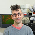
Ori Avinoam has a passion for understanding the molecular mechanisms of membrane remodeling. This began during his doctoral research under Professor Benjamin Podbilewicz at Technion—Israel Institute of Technology, where he identified and characterized fusogens in the nematode Caenorhabditis elegans, providing insights into cellular and viral fusogens. Ori then pursued a multidisciplinary postdoctoral fellowship at the European Molecular Biology Laboratory (EMBL) with Professor Marko Kaksonen and Professor John Briggs to study membrane remodeling during clathrin-mediated endocytosis. He joined the Weizmann Institute in 2017, where his lab has delved into the intricacies of membrane remodeling during skeletal muscle regeneration, the exocytosis of large secretory vesicles, and the fusion of extracellular vesicles and viruses. Adopting a holistic, interdisciplinary strategy combining genetics, biochemistry, and high-resolution imaging, his team has advanced our understanding of myogenesis, exocytosis, and extracellular vesicle fusion, providing a molecular and quantitative view on membrane remodeling in diverse contexts. Photo credit: Ohad Herches, Weizmann Institute of Science.
Lindsay Case

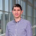
Lindsay Case received her PhD in cell biology at the University of North Carolina. During her graduate training, she worked with Clare Waterman at the National Institutes of Health to study integrin-based focal adhesions with superresolution fluorescence microscopy. She completed postdoctoral training in biophysics working with Michael Rosen at UT Southwestern Medical Center, where she investigated how phase separation promotes Arp2/3-dependent actin polymerization and developed a biochemical reconstitution of focal adhesions. She joined the MIT Biology Department as an Assistant Professor in 2021 and is currently studying how the spatial organization of molecules on the plasma membrane controls cell signaling. She aims to bridge biochemical and cell-based approaches to study cell adhesion, the cytoskeleton, and signaling. Photo credit: UT Southwestern.
Gautam Dey

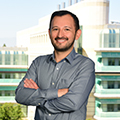
Gautam Dey started his group at EMBL in early 2021 after a postdoc in London working on the evolution of cell division with Buzz Baum. The group investigates the evolution of mitosis and nuclear remodeling using cell biology in multiple microbial model systems, comparative genomics, and experimental evolution. Gautam holds a PhD in systems biology from Stanford University and a research MSc from the National Centre for Biological Sciences in Bangalore, India. Gautam is an ERC Investigator (2023–2028) and a previous holder of a Marie-Sklodowska Curie postdoctoral fellowship (2017–2018) and a Stanford Graduate Fellowship in Science and Engineering (2009–2012). Photo credit: Kinga Lubowiecka, EMBL.
Stephanie Ellis

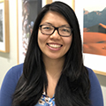
Stephanie Ellis studies the basic cellular and molecular principles that govern complex tissue assembly and homeostasis. She first got interested in this problem as a PhD student with Guy Tanentzapf at UBC, where she studied cell–ECM interactions in Drosophila embryogenesis. Then, as a postdoc with Elaine Fuchs at Rockefeller University, she set up model systems to study cell competition and fitness sensing in the developing mammalian skin. Now, at the University of Vienna, Stephanie and her group are uniting concepts and tools from cell and developmental biology, disease modeling, and biophysics to delineate conserved growth control mechanisms in heterogeneous tissues as they grow, reach their final size, and contend with threats to their homeostasis. Ultimately, work from the Ellis lab seeks to uncover fundamental insights pertaining to how cells within complex tissues communicate and are influenced by the phenotypes of their neighbors to ensure robust and healthy tissue function over long lifetimes. Photo credit: Luiza Puiu.
Elif Nur Firat-Karalar

Elif Nur Firat-Karalar studied molecular biology and genetics at Bilkent University, Turkey. She then moved to the University of California, Berkeley for her PhD work, where she investigated the mechanisms of actin nucleation under the supervision of Matthew Welch. During her postdoctoral work in the laboratory of Tim Stearns at Stanford University, she used proximity-mapping and biochemical approaches and identified the centriole proteome and interactome, revealing new mechanisms for centriole and cilium biogenesis. In 2014, Elif has established her laboratory, CytoLab, at Koç University in Turkey. Research in CytoLab focuses on studying the biology of the mammalian centrosome/cilium complex in health and in disease, with a particular focus on centriolar satellites. Elif is the recipient of two ERC Starting Grants, EMBO Young Investigator Award and TUBITAK National Leader Researcher Grant. Photo credit: Nurdan Usta.
Jonathan Friedman

Jonathan Friedman has a long-standing interest in the fundamental question of how organelles are spatially organized. Jonathan received his bachelor’s degree at Washington University in St. Louis. He performed his graduate work at the University of Colorado Boulder in Gia Voeltz’s lab, where he studied endoplasmic reticulum dynamics and inter-organelle contacts. As a postdoctoral fellow in Jodi Nunnari’s lab at the University of California, Davis, Jonathan examined mechanisms that control the internal architecture of mitochondria. He started his lab in 2018 at UT Southwestern Medical Center in the Department of Cell Biology. His laboratory studies how mitochondrial dynamics, functions, and quality control pathways are spatially regulated and modulated in the context of cellular metabolic demand. Photo credit: UT Southwestern.
Meng-meng Fu

Meng-meng completed her PhD from the University of Pennsylvania in 2013 and worked on axonal transport in the lab of Erika Holzbaur. She then started a postdoc at Stanford University in the lab of Ben Barres, working on microtubule regulation in oligodendrocytes. In 2020, she started her lab at the National Institute of Neurological Disorders and Stroke (NIH), working on the connection between liquid condensates in glia and neurological disease. In 2023, she moved to the University of California, Berkeley, where her lab currently works on cytoskeletal regulation, mRNA transport, and local translation in oligodendrocytes, astrocytes, and microglia.
Yaming Jiu

Yaming Jiu received her PhD in Biophysics from the Institute of Biophysics, Chinese Academy of Science, supervised by Professor Tao Xu, focusing on imaging techniques and membrane trafficking study. She continued with postdoc training in dynamic cytoskeletal remodeling in Professor Pekka Lappalainen’s laboratory in the University of Helsinki and was granted the title Docent in 2017. Since 2018, she has been leading the unit of cell biology and imaging study of pathogen host interaction as principal investigator in the Shanghai Institute of Immunity and Infection (formerly the Institut Pasteur of Shanghai), Chinese Academy of Sciences. Yaming Jiu’s current research interests lie in the field of the physiological and pathological functions of the dynamically regulated cytoskeleton in the context of tumor cell migration and pathogen infection (e.g., enteropathogenic bacteria, flavivirus), using various advanced imaging methods and in terms of in vivo complexity (e.g., composition of microbiota, ECM mechanics).
Anjali Kusumbe

Anjali Kusumbe pursued her doctoral studies with a fellowship from the Council of Scientific and Industrial Research, India, and was awarded a PhD in 2012. She then completed her postdoctoral research at the Max Planck Institute for Molecular Biomedicine, Germany, in 2016. Anjali is the head of the Tissue and Tumour Microenvironments Group at the MRC Weatherall Institute of Molecular Medicine, University of Oxford. She received an MRC Career Development Award in 2017 and an ERC Starting Grant in 2019 to lead her independent research program on vessel-tissue and vessel-immune interactions during tissue regeneration, aging, and metastasis. Anjali has received a number of awards for her work, including the British Society for Cell Biology Women in Cell Biology Award, the Royal Microscopical Society Life Sciences Medal, and the Rising Star Award from International Society for Regenerative Biology, among others.
Binyam Mogessie

Binyam Mogessie is an Assistant Professor of Molecular, Cellular and Developmental Biology and of Obstetrics, Gynecology, and Reproductive Sciences at Yale University. Originally from Ethiopia, Binyam received his BSc in biochemistry and cell biology in 2007 from Jacobs University Bremen in Germany and his PhD in Cell Biology in 2011 from the University of London. Binyam carried out his postdoc research at the MRC-LMB in Cambridge, UK and the Max Planck Institute for Biophysical Chemistry in Germany, where he discovered a function of the actin cytoskeleton in preventing aneuploidy in mammalian eggs. Binyam’s laboratory is dedicated to deciphering chromosome segregation with a special focus on unique cytoskeletal mechanisms that support healthy oocyte meiosis and embryo development. Photo credit: Yale University.
Pablo Lara-Gonzalez

Pablo Lara-Gonzalez is originally from Chile, where he graduated from the Universidad de Concepcion with a BSc in Biochemistry. He then moved to the UK where he obtained his PhD in Cell Biology at the University of Manchester, under the supervision of Dr. Stephen Taylor. Then, he moved to California to work as a postdoc in the laboratory of Arshad Desai at the Ludwig Institute for Cancer Research. Pablo is currently an assistant professor at the Department of Developmental and Cell Biology at the University of California, Irvine. Pablo’s research combines genetics with live-imaging microscopy, biochemistry, and proteomic approaches to study the molecular mechanisms that regulate cell cycle transitions and how these are regulated by developmental and environmental cues. Photo credit: University of California, Irvine.
Andrew Muroyama

Andrew Muroyama is an assistant professor in the Department of Cell and Developmental Biology at the University of California, San Diego. He received his BA from Swarthmore College and went on to complete his PhD with Dr. Terry Lechler at Duke University, where he studied the formation and function of non-centrosomal microtubules in mammalian epithelia. He conducted his postdoctoral research with Dr. Dominique Bergmann at Stanford University, exploring the mechanisms controlling asymmetric cell division in Arabidopsis thaliana. Since starting at UC San Diego in 2021, his group has focused on understanding how polarity within stem cell populations drives the formation of plant tissues. In particular, his lab is using quantitative imaging approaches to explore how the cytoskeleton regulates formation and maintenance of polarity across different length scales in developing plants.
Sonya Neal

Sonya Neal joined the faculty at the University of California, San Diego in 2018. She received her PhD in Molecular Biology from the University of California, Los Angeles for her work on understanding the mechanisms of mitochondria transport processes. She later pursued her postdoctoral studies in UC San Diego, where she studied the mechanistic actions of a widely conserved protein quality control pathway known as ER-associated degradation. In 2023, she became a Howard Hughes Medical Institute Freeman Hrabowski scholar. The Neal laboratory’s research focuses on the mechanisms of membrane protein and lipid homeostasis. Her lab is particularly interested in the physiological role of rhomboid-like proteins, which are intramembrane proteases and pseudoproteases known to function in removing substrates from the lipid bilayer to coordinate (1) signaling processes or (2) their degradation by the cytosolic ubiquitin proteasome system. Finally, her lab studies the role of rhomboid-like proteins in cancer and how their defects contribute to diseases resulting from malfunctions in protein/lipid homeostasis or signaling. They combine various approaches ranging from biochemistry, cell biology, and vertebrate biology to structural approaches to comprehensively understand rhomboid-like proteins. Photo credit: Gustavo Maselli.
Masayuki Onishi

Masayuki Onishi received his graduate training at the University of Tokyo, Japan, on a project that investigated the roles of the septin cytoskeleton during spore membrane morphogenesis in fission yeast. He then moved to Stanford University to work as a postdoc with Dr. John R. Pringle to study how yeast cells without a canonical contractile actomyosin ring still form a cleavage furrow. While still at Stanford, Masa launched a new project that investigated the mechanism of cytokinesis in the green alga Chlamydomonas reinhardtii. His current lab in the Department of Biology, Duke University studies how this unicellular relative of plants, which has no myosin-II, forms an animal-like cleavage furrow and how this process is coordinated with the division of the chloroplast, an organelle of bacterial origin.
Daniel Rios Barrera

Daniel Rios Barrera got his PhD in 2014 from the Universidad Nacional Autónoma de México (UNAM) Campus Juriquilla. He then moved to Germany, where he did his postdoctoral work at the European Molecular Biology Laboratory in Maria Leptin’s lab. Since 2020, he is a group leader at the Institute for Biomedical Research at UNAM in Mexico City. His team is interested in studying mechanisms of morphogenesis at multiple scales, from subcellular principles to whole tissue interactions. Specifically, the lab focuses on the development of the Drosophila tracheal system, an epithelium that exploits basic cellular machinery to build tubes and interacts with a range of different tissues to allow gas exchange. Current projects of the lab explore the role of vesicle trafficking in tracheal morphogenesis and the coordination between tracheal development and whole-embryo remodeling.
Samantha Stehbens

Samantha Stehbens is a cell biologist with an interest in understanding the fundamental mechanisms that regulate cell adhesion and the cytoskeleton. They completed their graduate studies at the University of Queensland with Professor Alpha Yap, where they uncovered how the microtubule cytoskeleton regulates cell–cell adhesion. Recognizing the importance of microscopy in their research, they made the strategic move to the University of California, San Francisco to work with Professor Torsten Wittmann, a cytoskeletal biologist renowned for live-cell spinning disc confocal microscopy. Samantha then discovered how microtubules coordinate protease secretion during migration to mediate cell–matrix adhesion disassembly. Returning to Australia, they applied their knowledge to investigate how cancer cells exploit fundamental mechanisms driving cell shape and motility to drive metastasis. Supported by an Australian Research Council Future Fellowship, they established their independent research program in 2020 at the University of Queensland. Their research group aims to understand how cells integrate secreted and biomechanical signals from their local microenvironment to facilitate movement and survival. They have uncovered a novel role for the microtubule cytoskeleton in protecting cells from rupture during 3D cell migration and invasion. Leveraging patient-derived tumor cells and advanced genetic and microfabrication techniques, they use state-of-the-art microscopy to explore cell migration mechanisms with the aim of improving strategies to prevent cancer metastasis. Photo credit: University of Queensland.


