We are pleased to introduce our newest members of the JCB editorial board. We are grateful to these and all of our board members for their contributions to JCB and service to the cell biology community.
Dominique Bergmann
Cell fate, stem cell behavior, and cell polarity in plants
Dominique Bergmann is a professor in the biology department at Stanford University, an investigator of the Howard Hughes Medical Institute, and an adjunct staff member of the Carnegie Institution, Department of Plant Biology. She has been fascinated by cell and developmental asymmetries since her PhD on early axis formation in C. elegans (Colorado University, Boulder, CO,with William B. Wood). As a postdoctoral researcher with Chris Somerville at the Carnegie Institution, she developed the Arabidopsis stomatal lineage (stomata are epidermal structures that regulate carbon dioxide and water exchange) into a model to understand how cells are specified to initiate asymmetric divisions, how asymmetric divisions are carried out in walled plant cells, and how the number and orientations of asymmetric divisions are dictated by the interplay of cell type–specific transcription factors, local peptide signaling, and global mechanical and environmental cues. Photo Courtesy of Ted Raab.
Magdalena Bezanilla
Actin cytoskeleton and plant cell growth
Magdalena Bezanilla is a professor of biology at the University of Massachusetts, Amherst. She completed a BS degree in physics at the University of California, Santa Barbara, and a PhD at Johns Hopkins University where she worked with Tom Pollard to characterize the role of myosin II in cytokinesis in fission yeast. She transitioned to plant cell biology as a postdoctoral fellow working with Ralph Quatrano at Washington University. Dr. Bezanilla has pioneered the use of the moss Physcomitrella patens as a model system to interrogate how proteins within the cell direct and regulate extracellular matrix deposition, ultimately affecting cell growth and morphogenesis. She has particularly focused on the regulation of the filamentous actin cytoskeletal network. The Bezanilla laboratory has developed such tools as RNA interference, quantitative complementation analyses, and rapid quantitative growth assays for use in P. patens to characterize cell growth and development. Recent work has led to the development of a working model for tip growth and cell division. Photo courtesy of the University of Massachusetts, Amherst.
Karlene Cimprich
Genome stability, DNA damage, and DNA replication
Karlene Cimprich is a professor in the Department of Chemical and Systems Biology at the Stanford University School of Medicine. Dr. Cimprich received her PhD in chemistry from Harvard University and remained there for postdoctoral work with Stuart Schreiber. Her lab focuses on understanding the mechanisms by which a cell maintains genome stability, particularly in the context of DNA damage and DNA replication. She identified the ATR checkpoint kinase as a postdoctoral fellow and much of her lab’s work focused on elucidating how its activation is linked to DNA replication. Her lab has also been instrumental in showing that the disruption of RNA processing is a prominent source of DNA damage in cells and determining the mechanism by which transcription-associated RNA–DNA hybrids and R-loops contribute to this damage. Dr. Cimprich is a AAAS fellow and a recipient of the Kimmel Scholar Award, Burroughs Wellcome New Investigator Award, and the Ellison Senior Scholar Award. Photo courtesy of Karlene Cimprich.
Judith Frydman
Molecular chaperones, protein folding, and degradation
Judith Frydman is Professor of Biology and Genetics at Stanford University School of Medicine. She is a member of Bio-X, the Stanford Cancer Institute, and the Stanford Neurosciences Institute, as well as a Faculty Fellow of Stanford ChEM-H. The Frydman lab uses multidisciplinary approaches to address fundamental questions about molecular chaperones, protein folding, and degradation, uncovering both basic mechanistic principles as well as identifying links to diseases such as cancer and neurodegeneration.
Yukiko Goda
Synaptic organization and plasticity
Yukiko Goda is a senior scientist at RIKEN Brain Science Institute. Research efforts in her group are directed towards delineating the mechanisms underlying the organization and plasticity of synapses and how the local synaptic interactions shape the properties of neural circuits in the mammalian brain. She received her PhD in biochemistry from Stanford University where she worked with Suzanne Pfeffer. After postdoctoral training in the laboratory of Chuck Stevens at the Salk Institute, she joined the faculty of the Division of Biology, University of California, San Diego, in 1997. She moved her group to the UK in 2002 as a senior group leader in the Medical Research Council Laboratory for Molecular Cell Biology at University College London, and then relocated to RIKEN Brain Science Institute in 2011 to take up her current position as a senior team leader and set up the Laboratory for Synaptic Plasticity and Connectivity. Photo courtesy of RIKEN Brain Science Institute.
Bruce Goode
Cytoskeleton in cell motility, cell morphogenesis, and intracellular transport
Bruce Goode is a professor of biology at Brandeis University. Dr. Goode earned a BS in biology from the University of California, Santa Barbara, received his PhD at the same institution working in the laboratory of Stuart Feinstein, and conducted his postdoctoral research in the laboratories of David Drubin and Georjana Barnes at the University of California, Berkeley. In 2000, Dr. Goode started his lab at Brandeis University, where his research focuses on dynamic rearrangements of the actin and microtubule cytoskeletons that drive cell motility, cell morphogenesis, and intracellular transport. Specific areas of interest include defining mechanisms of actin filament assembly, actin filament disassembly and turnover, and coordination of microtubule and actin dynamics. Work is equally divided between yeast and mammalian systems and is multidisciplinary in nature, combining in vitro single molecule total internal relection fluorescence imaging, genetics, biochemistry, and live cell imaging. Dr. Goode has received scholar awards from the Pew Charitable Trust, March of Dimes, and American Cancer Society, and a Research Career Development Award from the National Institutes of Health. Dr. Goode is on the F1000 advisory board and was Editor-in-Chief of Cytoskeleton from 2009 to 2016. Photo courtesy of Bruce Goode.
Roger Greenberg
DNA repair, ubiquitin signaling, and cancer
Roger Greenberg is an associate professor in the Department of Cancer Biology at the University of Pennsylvania, where he also serves as Director of Basic Science for the Basser Center for BRCA. He received his MD and PhD from the Albert Einstein College of Medicine. Performing thesis studies with Professor Ronald DePinho, Dr. Greenberg demonstrated the first evidence that telomere shortening suppresses carcinogenesis in vivo. Subsequently, Dr. Greenberg completed postdoctoral training at the Dana-Farber Cancer Institute with Professor David Livingston, where he defined a BRCA1-centered tumor suppressor network. His independent laboratory investigates basic mechanisms of genome integrity maintenance and their impact on cancer etiology and response to therapy. His group discovered that ubiquitin chains serve as a platform for BRCA1 DNA damage recognition, identified biallelic mutations in BRCA1 as a cause of Fanconi anemia, and developed novel cellular systems that enabled his group to discover ATM kinase–dependent transcriptional silencing near DNA double-strand breaks and delineate homologous recombination–dependent alternative telomere lengthening mechanisms in cancer. Photo courtesy of Daniel Burke.
James Hurley
Structural membrane biology
James Hurley is the Judy C. Webb Chair and Professor of Biochemistry, Biophysics, and Structural Biology at the University of California, Berkeley. He obtained his PhD at the University of California, San Francisco, and was a postdoctoral fellow at the University of Oregon. He was an investigator at the Laboratory of Molecular Biology (LMB) at the National Institute of Diabetes and Digestive and Kidney Diseases, National Institutes of Health, and Chief of the Section on Structural Biology and Cell Signalling at LMB from 1998 to 2013. The Hurley lab studies interactions between proteins and membrane lipids; their roles in autophagy, membrane scission, and coated vesicle formation; and how pathogens such as HIV subvert these processes. The Hurley lab uncovers the molecular mechanism behind these interactions using interdisciplinary approaches, including cryoelectron microscopy, x-ray crystallography, biochemical reconstitution, and live-cell imaging. Dr. Hurley received the Hans Neurath Award in 2014 from the Protein Society and the Outstanding Science Award from SER-CAT in 2009. Photo courtesy of Brittany Hosea-Small, University of California, Berkeley.
Satyajit Mayor
Membrane organization and endocytosis
Satyajit “Jitu” Mayor has taken a multidisciplinary approach combining cell biology with physics and chemistry to study the organization and endocytic trafficking of membrane lipids, transmembrane and lipid-anchored proteins in membranes of living cells. The trajectory of this work has led him to explore the fine structure of the plasma membrane, combining ideas from soft-matter and membrane biophysics. He also uses tools of molecular genetics to explore the implications of these findings in the construction of signaling platforms and endocytic pathways, and the roles they may play in building up tissue architectures. Dr. Mayor studied Chemistry at the Indian Institute of Technology, Mumbai, and obtained his PhD in life sciences from the Rockefeller University. He was a postdoctoral fellow at Columbia University, where he developed tools to study the trafficking of membrane lipids and glycosylphosphatidylinositol-anchored proteins in mammalian cells using quantitative fluorescence microscopy. Currently he is Senior Professor and Director at National Centre for Biological Sciences (NCBS) Tata Institute for Fundamental Research and the Institute for Stem Cell Biology and Regenerative Medicine in Bangalore, India. He is a fellow of the Indian Academy of Sciences and National Academy of Sciences, and a foreign associate of the US National Academy of Sciences and European Molecular Biology Organization. He is a recipient of several national and international awards including the Shanti Swarup Bhatnagar Award, the World Academy Prize for Biology, and the Infosys Prize for Life Sciences. Photo courtesy of NCBS.
Ewa Paluch
Cellular morphogenesis
Ewa Paluch is Professor of Cell Biophysics and a Medical Research Council (MRC) program leader at the MRC Laboratory for Molecular Cell Biology, University College London. Dr. Paluch studied physics and mathematics at the École Normale Supérieure in Lyon, France. She did her PhD (2001–2005) at the Curie Institute in Paris, under the supervision of Cécile Sykes and Michel Bornens, investigating actin network mechanics in vitro and in cells. In 2006, she started her own research group at the Max Planck Institute of Molecular Cell Biology and Genetics in Dresden, Germany, as a joint appointment with the International Institute of Molecular and Cell Biology in Warsaw. She moved her laboratory to University College London in 2013. Since 2014, she has also been the head of the Institute for the Physics of Living Systems, a cross-faculty initiative aiming to promote collaborations between physicists and biologists at University College London. She received a Philip Leverhulme Prize in Biological Sciences in 2014 and the Hooke Medal from the British Society for Cell Biology in 2017. Dr. Paluch’s laboratory studies the mechanisms controlling cellular morphogenesis. Since cell shape is ultimately defined by cellular mechanical properties and by the cell’s physical interactions with its environment, biophysical approaches are essential to understand cell shape control. The lab combines cell biology, biophysics, and quantitative imaging, and works in close collaboration with theoretical physicists, to investigate cell shape regulation. They particularly focus on the cellular actin cortex, a thin cytoskeletal network that underlies the plasma membrane and drives most shape changes in animal cells. Photo courtesy of Kostas Margitudis, MPI-CBG, Dresden, Germany.
Billy Tsai
Host–pathogen interactions, ER protein quality control, and viral infection
Billy Tsai is currently the Corydon Ford Professor of the cell and developmental biology department at the University of Michigan Medical School. He received his BA and MS from University of California, Los Angeles, received a PhD from Harvard University, and was a Damon-Runyon postdoctoral fellow in the laboratory of Dr. Tom Rapoport at Harvard Medical School. Dr. Tsai was the recipient of the Burroughs Wellcome Fund Investigators in Pathogenesis of Infectious Disease award. His laboratory is centrally focused on clarifying the molecular basis by which polyomavirus and cholera toxin penetrate the ER membrane to cause disease. In addition, his laboratory studies how ER quality control processes control the fate of proinsulin mutants to impact diabetes. Photo courtesy of the University of Michigan.
Bas van Steensel
Architecture and functions of chromosomes and chromatin
Bas van Steensel obtained his PhD at the University of Amsterdam in 1995. He was a postdoctoral fellow in the lab of Titia de Lange (Rockefeller University) and subsequently with Steven Henikoff (Fred Hutchinson Cancer Research Center). Since 2001 he leads a research group at the Netherlands Cancer Institute in Amsterdam. His lab develops and applies new methods to study the structure and composition of chromatin and chromosomes, the spatial organization of the genome inside the nucleus, and mechanisms of gene regulation. Photo courtesy of Bas van Steensel.
Mark von Zastrow
G protein–coupled receptors, membrane trafficking, and receptor-mediated signaling
Mark von Zastrow is a professor in the departments of psychiatry and cellular and molecular pharmacology at the University of Caliornia, San Francisco. He obtained his PhD with J. David Castle and George Palade at Yale University and did postdoctoral work with Jack Barchas and Brian Kobilka at Stanford University. His research group investigates relationships between membrane trafficking and receptor-mediated signaling processes, focusing primarily on G protein–coupled receptors. He is particularly interested in understanding the cellular basis of neuromodulation, brain disease, and receptor-mediated drug action. Photo courtesy of Mark von Zastrow.
Xiaochen Wang
Phagocytosis and lysosome homeostasis
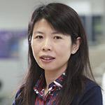
Xiaochen Wang is a principal investigator at the Institute of Biophysics (IBP), Chinese Academy of Sciences (CAS). Using combinatory approaches of genetics, cell biology, and biochemistry, Wang’s lab studies mechanisms controlling various aspects of apoptotic cell removal including recognition, internalization, and degradation of cell corpses in the model organism Caenorhabditis elegans. More recently, Wang’s lab has been developing and applying C. elegans as a multicellular genetic model for systematic investigation of lysosome homeostasis, with the aim of identifying signals/cellular processes that trigger or are associated with diverse lysosomal changes, dissecting the underlying regulatory mechanisms and revealing their physiological significance. Dr. Wang received her PhD in molecular biology from Peking University and completed postdoctoral work at the University of Colorado, Boulder. She was an assistant investigator from 2006 and associate investigator from 2011 at the National Institute of Biological Sciences (Beijing, China) until she moved to IBP, CAS, in 2016. Dr. Wang was a Howard Hughes Medical Institute International Early Career Scientist. Photo courtesy of China Young Women in Science Fellowships.
Hong Zhang
Autophagy
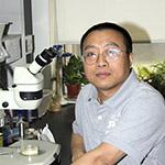
Hong Zhang is an investigator in the Institute of Biophysics, Chinese Academy of Sciences (CAS). He graduated from Albert Einstein College of Medicine and did his postdoctoral training with Dr. Daniel Haber in the Massachusetts General Hospital Cancer Center, Harvard Medical School. Before joining the Institute of Biophysics, CAS, in 2012, he was an assistant investigator (2004–2009) and associate investigator (2009–2012) of the National Institute of Biological Sciences, Beijing. Zhang’s lab demonstrated that during Caenorhabditis elegans embryogenesis, components of specialized protein aggregates, called P granules, are removed by autophagy in somatic cells. Using this as a model, his lab performed the first systematic genetic screens for novel autophagy genes in higher eukaryotes, resulting in identification of a set of metazoan-specific autophagy genes, known as epg genes. His lab is currently investigating whether different cells and tissues in multicellular organisms contain specific variants of the autophagy machinery and how autophagy activity is coordinately regulated to maintain cell, tissue, and organism homeostasis. He was a Howard Hughes Medical Institute International Early Career Scientist and has received the sixth C.C.Tan (Jia-Zhen Tan) Life Science Award. Photo courtesy of Jianwei Ni.


