



Many cancers use an alternative lengthening of telomeres (ALT) pathway for telomere maintenance. ALT telomeric DNA synthesis occurs in ALT-associated PML bodies (APBs). However, the mechanisms by which APBs form are not well understood. Here, we monitored the formation of APBs with time-lapse imaging employing CRISPR knock-in to track the promyelocytic leukemia (PML) protein at endogenous levels. We found APBs form via two pathways: telomeres recruit PML proteins to nucleate PML bodies de novo, or telomeres fuse with preformed PML bodies. Both nucleation and fusion of APBs require interactions between SUMO and SUMO interaction motifs (SIMs). Moreover, APB nucleation is associated with higher levels of SUMO and SUMO-mediated recruitment of DNA helicase BLM, resulting in more robust telomeric DNA synthesis. Finally, further boosting SUMO levels at telomeres enhances APB nucleation, BLM enrichment, and telomeric DNA synthesis. Thus, high SUMO levels at telomeres promote APB nucleation and stronger ALT activity.
Introduction
Cancer cells need to overcome replication-associated telomere shortening to maintain proliferation (Okamoto and Seimiya, 2019). While most cancers achieve this via reactivating telomerase (Shay, 2016), ∼15% of cancers utilize alternative lengthening of telomeres (ALT) through a recombination-based repair mechanism called break-induced replication (BIR) (Pickett and Reddel, 2015; Zhang and Zou, 2020; Dilley et al., 2016). ALT takes place in specialized promyelocytic leukemia (PML) nuclear bodies (PML-NBs) called ALT-associated PML-NBs (APBs) that enrich both clustered telomeres and BIR proteins, thus serving as a hub for BIR-mediated telomere recombination (Brouwer et al., 2009; Chung et al., 2011; Zhang et al., 2019; Draskovic et al., 2009). APBs have provided a main ALT diagnostic tool, and their reduced formation can potentially halt ALT cancer proliferation (Zhao et al., 2024).
SUMOylation, the process by which the small ubiquitin modifier (SUMO) is conjugated to target proteins, has been shown to play a key role in APB formation and ALT (Chung et al., 2012; Osterwald et al., 2015; Min et al., 2019). SUMOylation inhibitors can reduce APB levels and ALT cell growth, suggesting their therapeutic potentials against ALT cancers (Zhao et al., 2024). SUMOylation affects three groups of proteins important to APB formation and ALT. First, SUMOylation of telomeric proteins, such as telomere repeat factor 1 (TRF1), TRF2, TIN2, and RAP1, is required for APB formation (Brouwer et al., 2009; Chung et al., 2011; Potts and Yu, 2007). Second, SUMOylation of DNA repair factors, such as Rad52 and BLM, enables their recruitment to APBs (Zhang et al., 2021). Third, SUMOylation of the PML protein, the scaffold of the PML-NBs, is critical for PML-NB and APB formation (Chung et al., 2011; Duprez et al., 1999; Tatham et al., 2008; Zhao et al., 2024). A prominent role of SUMOylation is to promote interactions among telomeric proteins, DNA repair proteins, and the PML protein via interactions between the SUMO moieties and SUMO interaction motifs (SIMs). Numerous SIMs have been found on these proteins, and their SUMOylation at many sites permits multivalent SUMO–SIM interactions, which can drive phase separation to form liquid condensates (Keiten-Schmitz et al., 2021; Raman et al., 2013; Shen et al., 2006; Zhang et al., 2020). Indeed, the importance of SIMs in APB functions is demonstrated as SIMs of DNA repair factors, such as Rad51AP1 and BLM, are required for their APB localization (Loe et al., 2020; Min et al., 2019). In addition, the SIM in the PML protein is key for PML-NB and APB formation (Shen et al., 2006; Lallemand-Breitenbach and Thé, 2010; Zhang et al., 2021).
While the requirement of SUMOylation for APB formation is well established, important details are lacking regarding APB formation pathways and the roles of SUMOylation in this process. It is unclear whether APBs form via de novo nucleation of PML-NBs on telomeres or via recruiting telomeres to preexisting PML-NBs. In addition, it is unknown how different SUMO paralogs and SUMO ligases affect APB formation pathways and if they do so by controlling SUMOylation of specific proteins. Addressing these questions requires live-cell time-lapse imaging of APBs in a condition wherein new APB formation can be induced and monitored in real time. In addition, the time-lapse imaging must be done at the endogenous PML protein level, as overexpressing PML is known to affect PML-NB formation (Sahin et al., 2014; Corpet et al., 2020). One method that allows inducible APB formation is the TRF1–FokI system (Cho et al., 2014; Dilley et al., 2016). In this system, FokI is fused with the TRF1, a destabilization domain (DD), and a modified estrogen receptor (ER) domain. The addition of 4-hydroxytamoxifen and shield1 induces FokI nuclease release to nucleoplasm and translocation to telomeres. Telomere localized FokI–TRF1 then triggers DNA breaks exclusively at telomeres, leading to the formation of APBs with all endogenous APB characteristics, such as telomere clustering and telomeric DNA synthesis. Another method for APB formation with temporal control entails tethering SUMO or SIM to telomeres using a chemical-induced protein-dimerization system (Lackner et al., 2022; Zhao et al., 2021). In this system, the dimerizer trimethoprim (TMP)-fluorobenzamide-Halo ligand (TFH) is composed of chemically linked TMP and Halo ligand that can interact with the Escherichia coli dihydrofolate reductase (eDHFR) and the Halo-enzyme, respectively. Adding the dimerizer to cells expressing SUMO/SIM fused to eDHFR and TRF1 fused to Halo-enzyme will target SUMO/SIM to telomeres to form APBs on demand. This strategy can be coupled with live-cell time-lapse imaging and prevents the toxicity caused by constitutive telomere targeting. Importantly, we engineered an ALT-positive cell line, U2OS, so that the endogenous PML protein is tagged with the fluorescent protein Clover (Pinder et al., 2015), allowing us to follow APB formation without overexpressing the PML protein.
Taking advantage of the TRF1–FokI system, the SUMO/SIM telomere targeting method, and the PML-Clover U2OS cell line, we monitored APB formation dynamics using live-cell time-lapse imaging. We examined two possible pathways for APB formation. First, given that the PML protein is decorated with SIMs and conjugated with SUMO moieties (Kamitani et al., 1998; Shen et al., 2006), it may be recruited to telomeres enriched with SUMO/SIM via SUMO–SIM interactions to form PML-NBs on telomeres, and thus APBs. This model of de novo formation of APBs via nucleating PML-NBs on telomeres is referred to as the APB nucleation model (Fig. 1 A top). Alternatively, considering that SUMO mediates phase separation of DNA repair factors located on ALT telomeres without PML (Zhao et al., 2024), the thus-formed condensates may fuse with preexisting PML-NBs that are also condensates enriched with SUMO to form APBs. We refer to this hypothesis, entailing two types of condensates coalescing, as the APB fusion model (Fig. 1 A bottom). We provide evidence that both mechanisms are present in the same cell and both require SUMO–SIM interactions. We detail the contributions of SUMO/SIM from the PML protein, different SUMO paralogs, and SUMO ligases for APB formation and ALT telomeric DNA synthesis. We report a set of observations indicating that SUMO levels at telomeres influence the two APB formation mechanisms. First, we find that APB nucleation occurs more often on telomeres with higher SUMO levels compared with APB fusion. Second, boosting the telomeric SUMO level can promote more APB nucleation. Third, APB nucleation is more sensitive to reducing telomere SUMO levels. These data are consistent with each other and indicate that SUMO level is a determining factor in the choices of APB formation mechanisms. Finally, we show the functional difference of these two APB formation mechanisms, with APB nucleation leading to more robust telomere synthesis. Collectively, our data provide direct visualization of two APB formation routes, reveal differential ALT potentials of these two pathways, and uncover how the two APB formation choices depend on SUMO levels at telomeres.

APBs are formed by both fusion and nucleation. (A) Schematics of the APB nucleation and fusion model. SUMOylation occurs on telomere-binding proteins and DNA repair factors (Zhang et al., 2020). For simplicity, these proteins are not depicted in the schematic. (B) Top: Schematics for chemical-induced targeting of mCherry-FokI to telomeres. The nuclease FokI was fused with the TRF1, destabilization domain (DD), and ER (modified estrogen receptor) domain. The addition of 4-hydroxyestradiol (4-OHT, a selective estrogen receptor modulator that binds to ER) and shield1 (stabilizes the protein tagged with the DD domain) allows FokI fused to mCherry (mCh) and the telomeric TRF1 protein to be released into the nucleoplasm and localized to telomeres, inducing telomere-specific DNA breaks, protein SUMOylation, and APB formation. Middle: Representative images of U2OS PML-Clover cells expressing mCh-TRF1–FokI with the treatment of 4-OHT and shield1 to induce DNA damage after the first time frame. Yellow arrows indicate APBs. Bottom: Examples of APB formation by fusion and nucleation after FokI induction, 5 min between images. (C) Quantification of APB and PML-NB numbers per cell after FokI induction. The numbers were normalized by the values at the first time point for each cell, >16 cells per group, three independent experiments. (D) Quantification of the number of newly formed APBs per cell through nucleation and fusion after inducing FokI or dimerizing SUMO1/3 (nonconjugable)/SIM for 2 h. More than 30 cells per group, three independent experiments. (E) The integrated intensity of TRF1 foci (left) and the time for APB formation (right) via nucleation and fusion after inducing FokI or dimerizing SUMO3 (nonconjugable) for 2 h. Each dot represents one cell, three independent experiments, >42 cells per group. (F) Top: Schematics for TFH-induced protein targeting to telomeres via a dimerization strategy. SUMO or other proteins can be fused to mCherry and eDHFR, while TRF1 is fused to the 3xHalo enzyme. The dimerizer, TFH, can interact with eDHFR and the Halo enzyme to dimerize SUMO or other proteins fused to mCherry-eDHFR with TRF1-3Halo, thus targeting them to telomeres. Middle: Representative images of U2OS PML-Clover cells expressing mCh-eDHFR-SUMO3 (nonconjugable) and 3xHalo-TRF1 with the addition of the dimerizer at indicated time points. Yellow arrows indicate APBs. Bottom: Examples of APB formation by fusion and nucleation after dimerizing SUMO3 (nonconjugable), 5 min between images. (G) Quantification of APB and PML-NB numbers per cell after dimerizing SUMO3 (nonconjugable) (left), SUMO3(FAAA) (middle), and SIM (right) to PML-Clover cells. The numbers were normalized based on the count for each cell at the first time point. More than 16 cells per group, three independent experiments. Scale bars, 5 or 1 μm for the zoomed-in images.
APBs are formed by both fusion and nucleation. (A) Schematics of the APB nucleation and fusion model. SUMOylation occurs on telomere-binding proteins and DNA repair factors (Zhang et al., 2020). For simplicity, these proteins are not depicted in the schematic. (B) Top: Schematics for chemical-induced targeting of mCherry-FokI to telomeres. The nuclease FokI was fused with the TRF1, destabilization domain (DD), and ER (modified estrogen receptor) domain. The addition of 4-hydroxyestradiol (4-OHT, a selective estrogen receptor modulator that binds to ER) and shield1 (stabilizes the protein tagged with the DD domain) allows FokI fused to mCherry (mCh) and the telomeric TRF1 protein to be released into the nucleoplasm and localized to telomeres, inducing telomere-specific DNA breaks, protein SUMOylation, and APB formation. Middle: Representative images of U2OS PML-Clover cells expressing mCh-TRF1–FokI with the treatment of 4-OHT and shield1 to induce DNA damage after the first time frame. Yellow arrows indicate APBs. Bottom: Examples of APB formation by fusion and nucleation after FokI induction, 5 min between images. (C) Quantification of APB and PML-NB numbers per cell after FokI induction. The numbers were normalized by the values at the first time point for each cell, >16 cells per group, three independent experiments. (D) Quantification of the number of newly formed APBs per cell through nucleation and fusion after inducing FokI or dimerizing SUMO1/3 (nonconjugable)/SIM for 2 h. More than 30 cells per group, three independent experiments. (E) The integrated intensity of TRF1 foci (left) and the time for APB formation (right) via nucleation and fusion after inducing FokI or dimerizing SUMO3 (nonconjugable) for 2 h. Each dot represents one cell, three independent experiments, >42 cells per group. (F) Top: Schematics for TFH-induced protein targeting to telomeres via a dimerization strategy. SUMO or other proteins can be fused to mCherry and eDHFR, while TRF1 is fused to the 3xHalo enzyme. The dimerizer, TFH, can interact with eDHFR and the Halo enzyme to dimerize SUMO or other proteins fused to mCherry-eDHFR with TRF1-3Halo, thus targeting them to telomeres. Middle: Representative images of U2OS PML-Clover cells expressing mCh-eDHFR-SUMO3 (nonconjugable) and 3xHalo-TRF1 with the addition of the dimerizer at indicated time points. Yellow arrows indicate APBs. Bottom: Examples of APB formation by fusion and nucleation after dimerizing SUMO3 (nonconjugable), 5 min between images. (G) Quantification of APB and PML-NB numbers per cell after dimerizing SUMO3 (nonconjugable) (left), SUMO3(FAAA) (middle), and SIM (right) to PML-Clover cells. The numbers were normalized based on the count for each cell at the first time point. More than 16 cells per group, three independent experiments. Scale bars, 5 or 1 μm for the zoomed-in images.
Results
Telomere breaks induce APB formation via both nucleation and fusion
We used the TRF1–FokI system summarized above to induce APB formation in the PML-Clover U2OS cell line and followed the dynamics of newly formed APBs using time-lapse imaging (Fig. 1 B top). Since PML is the major structural component of PML-NBs, PML-Clover–enriched foci represent PML-NBs (Pinder et al., 2015). Accordingly, PML-Clover foci that are enriched in mCherry-TRF1–FokI represent APBs. Live imaging showed an increase in the number of APBs over time (Fig. 1 B middle; Video 1), indicating that new APBs were formed after inducing FokI. As a control, inducing the enzymatically dead mutant of FokI (D450A) failed to increase APB numbers (Fig. S1 A), confirming that new APB formation was induced by telomeric-specific DNA breaks as seen previously (Cho et al., 2014).
Inducing DNA damage at telomeres in PML-Clover cells. Movie for Fig. 1 B. Left: Composite of FokI–TRF1 (magenta) and PML (green); middle: mCh-FokI-TRF1; right: PML. 4-OHT and shield1 were added to cells after the first time point to induce damage. The boxes show APBs formed by fusion and nucleation. Scale bars, 5 μm. 4-OHT, 4-hydroxyestradiol.
Inducing DNA damage at telomeres in PML-Clover cells. Movie for Fig. 1 B. Left: Composite of FokI–TRF1 (magenta) and PML (green); middle: mCh-FokI-TRF1; right: PML. 4-OHT and shield1 were added to cells after the first time point to induce damage. The boxes show APBs formed by fusion and nucleation. Scale bars, 5 μm. 4-OHT, 4-hydroxyestradiol.

Role of SUMO and SIM in APB formation. (A) Representative images (left) and quantification (right) of APB and PML-NB numbers in PML-Clover U2OS cells expressing mCh-TRF1–FokI D450A with the treatment of 4-hydroxyestradiol (4-OHT) and shield1 to induce FokI after the first time frame. Numbers were normalized to the value at the first time point for each cell, >17 cells per group, three independent experiments. (B) Representative images and quantification of APBs in PML-Clover U2OS cells expressing 3xHalo-TRF1 with SUMO1/3 (WT) after adding the dimerizer for 2 h. More than 22 cells per group, three independent experiments. (C) Representative images of U2OS cells, PML-Clover U2OS cells expressing 3xHalo-TRF1 with mCh-eDHFR-SUMO3(FAAA) (left), mCh-eDHFR-SIM (middle), and mCh-eDHFR-SIM(VADA) (right) after adding the dimerizer following the first time point. Yellow arrows indicate APBs. (D) Quantification of APB and PML-NB numbers per cell after recruiting SIM(VADA) to PML-Clover telomeres. Numbers were normalized to the value at the first time point for each cell, >14 cells per group, three independent experiments. (E) Representative images of U2OS PML KO cells expressing 3xHalo-TRF1, PML (SUMO-) (left and middle) or PML (SIM-) (right), and mCh-eDHFR-SUMO3 (nonconjugable) (left) or mCh-eDHFR-SIM (middle and right) after adding the dimerizer after the first time frame. (F) Quantification of APB and PML-NB numbers after recruiting SUMO3 (nonconjugable)/SIM in PML KO cells expressing PML(SIM-) or (SUMO-). Numbers were normalized to the value at the first time point for each cell, >17 cells per group, three independent experiments. (G) Representative images (left), quantification of SUMO1 localization on telomeres (middle), and quantification of APB number (right) in G2-arrested U2OS PML KO cells after expressing PML WT, PML(SUMO-), or PML(SIM-). Each dot represents one cell, three independent experiments; >45 cells per group. Scale bars, 5 μm.

Role of SUMO and SIM in APB formation. (A) Representative images (left) and quantification (right) of APB and PML-NB numbers in PML-Clover U2OS cells expressing mCh-TRF1–FokI D450A with the treatment of 4-hydroxyestradiol (4-OHT) and shield1 to induce FokI after the first time frame. Numbers were normalized to the value at the first time point for each cell, >17 cells per group, three independent experiments. (B) Representative images and quantification of APBs in PML-Clover U2OS cells expressing 3xHalo-TRF1 with SUMO1/3 (WT) after adding the dimerizer for 2 h. More than 22 cells per group, three independent experiments. (C) Representative images of U2OS cells, PML-Clover U2OS cells expressing 3xHalo-TRF1 with mCh-eDHFR-SUMO3(FAAA) (left), mCh-eDHFR-SIM (middle), and mCh-eDHFR-SIM(VADA) (right) after adding the dimerizer following the first time point. Yellow arrows indicate APBs. (D) Quantification of APB and PML-NB numbers per cell after recruiting SIM(VADA) to PML-Clover telomeres. Numbers were normalized to the value at the first time point for each cell, >14 cells per group, three independent experiments. (E) Representative images of U2OS PML KO cells expressing 3xHalo-TRF1, PML (SUMO-) (left and middle) or PML (SIM-) (right), and mCh-eDHFR-SUMO3 (nonconjugable) (left) or mCh-eDHFR-SIM (middle and right) after adding the dimerizer after the first time frame. (F) Quantification of APB and PML-NB numbers after recruiting SUMO3 (nonconjugable)/SIM in PML KO cells expressing PML(SIM-) or (SUMO-). Numbers were normalized to the value at the first time point for each cell, >17 cells per group, three independent experiments. (G) Representative images (left), quantification of SUMO1 localization on telomeres (middle), and quantification of APB number (right) in G2-arrested U2OS PML KO cells after expressing PML WT, PML(SUMO-), or PML(SIM-). Each dot represents one cell, three independent experiments; >45 cells per group. Scale bars, 5 μm.
Tracing new APB formation after telomere break induction revealed both fusion and nucleation events. We define fusion events as the collision of FokI–TRF1-marked telomere foci with the preexisting PML foci, which resulted in the sudden appearance of APBs enriched in both PML and TRF1 (Fig. 1 B bottom left; Video 1). We define nucleation events as a gradual gain of PML signals on telomeres without a collision of preexisting PML foci with telomere foci (Fig. 1 B bottom right; Video 1). To confirm the visual inspection, we quantified the change in the total number of PML-NBs over time. We expect the PML-NB number to remain unchanged for APB formation via fusion because it is simply a re-localization of existing PML-NBs to telomeres. On the other hand, we anticipate the PML-NB number to increase for APB formation via nucleation because new PML-NBs are formed on telomeres. Confirming that APBs are formed by nucleation after TRF1–FokI induction, we observed an increase in the PML-NB number (Fig. 1 C), even though photobleaching could potentially reduce PML-NB numbers. The increase in PML-NB number was not seen when using the TRF1–FokI (D450A) construct as expected (Fig. S1 A).
Quantifying the number of APBs formed via fusion versus nucleation revealed that fusion events were about twice as frequent compared with nucleation events during the 2 h following FokI induction (Fig. 1 D). Overall, telomeres that nucleated APBs had a higher intensity than those engaged in APB formation via fusion (Fig. 1 E left). Quantifying the time of APB formation revealed that nucleation of APBs happened faster than fusion (Fig. 1 E right). While the underlying reason for this difference is unclear, a simple interpretation is that PML proteins diffuse faster than PML-NBs in the nucleoplasm. These results show that APBs can form via nucleation and fusion in the same cell, but these pathways differ in terms of speed and frequency as well as the level of telomere signals. We conclude that in the context of telomere breaks, APB fusion occurs more frequently, while APB nucleation happens faster and is associated with stronger telomeric signals.
Targeting SUMO to telomeres potently induces APB nucleation and fusion
To understand the dependence of the two APB formation pathways on SUMO, we used the SUMO–telomere targeting system capable of inducing new APBs in the PML-Clover cells (Fig. 1 F top). We examined SUMO3, one of the three human SUMO paralogs, focusing on its nonconjugable form (C-terminal di-Gly motif mutated to di-Val) to prevent conjugation (Banani et al., 2016). We found that recruiting SUMO3 to telomeres resulted in APB formation via both nucleation and fusion, as shown by live-imaging data and quantifications of APB and PML-NB numbers over time (Fig. 1, D and F middle, bottom; Fig. 1 G left; Video 2). As seen in the TRF1–FokI system, the SUMO targeting systems also show that APB nucleation was associated with stronger telomere signals (Fig. 1 E left) and occurred faster than fusion (Fig. 1 E right). SUMO targeting induced APB formation primarily through nucleation and at a level greater than FokI induction, suggesting that SUMO enrichment at telomeres promotes APB formation via nucleation.
Recruiting SUMO3 to telomeres in PML-Clover cells. 3xHalo-TRF1 was co-transfected. Movie for Fig. 1 F. Left: Composite of SUMO3 (magenta) and PML (green); middle: mCh-eDHFR-SUMO3; right: PML. The dimerizer was added to cells after the first time point to induce dimerization. The box shows APBs formed by fusion and nucleation. Scale bars, 5 μm.
Recruiting SUMO3 to telomeres in PML-Clover cells. 3xHalo-TRF1 was co-transfected. Movie for Fig. 1 F. Left: Composite of SUMO3 (magenta) and PML (green); middle: mCh-eDHFR-SUMO3; right: PML. The dimerizer was added to cells after the first time point to induce dimerization. The box shows APBs formed by fusion and nucleation. Scale bars, 5 μm.
Dimerizing the WT SUMO3 also formed APBs via both nucleation and fusion, but at a level higher than dimerizing the nonconjugable form of SUMO3 (Fig. S1 B). Targeting SUMO1 to telomeres led to outcomes similar to those described for SUMO3 targeting, for both WT and nonconjugable SUMOs (Fig. 1 D and Fig. S1 B). This suggests that the level of SUMO, rather than the identity of SUMO paralogs, is more important in determining APB formation via nucleation or fusion. Collectively, these data indicate that either telomere breaks or SUMO–telomere tethering resulted in new APB formation through fusion and nucleation, with nucleation being the faster pathway. The observed differences between the two APB-inducing systems can be unified by the theory that APB nucleation is favored at telomeres with higher SUMO levels.
Both pathways of APB formation require SUMO–SIM interactions
As both APB nucleation and fusion can occur upon SUMO tethering at telomeres, we asked whether the role of SUMO in these processes requires its binding to SIM. To this end, we recruited SUMO1 or SUMO3 mutants that could not interact with SIM (FKVK in SUMO1 and FKIK in SUMO3 mutated to FAAA) to telomeres (Banani et al., 2016). We found reduced APB formation via both nucleation and fusion compared with their WT counterparts (Fig. 1 G middle; Fig. S1 C left; Video 3). Nucleation decreased more dramatically than fusion for both mutants (Fig. 1 D), indicating that nucleation of APBs relies more heavily on SUMO–SIM interactions at the telomere compared with fusion. A similar conclusion was reached when examining telomere targeting of SIM or its SUMO-binding mutant (VIDL mutated to VADA) (Banani et al., 2016), since recruiting SIM, but not its mutant, led to APB formation via nucleation and fusion (Fig. 1 D and Video 4; Fig. S1 C middle, right, Fig. S1 D). Collectively, these results support the notion that SUMO–SIM interactions are critical for both APB formation pathways.
Recruiting SUMO3(FAAA) to telomeres in PML-Clover cells. 3xHalo-TRF1 was co-transfected. Movie for Fig. S1 C. Left: Composite of SUMO3(FAAA) (magenta) and PML (green); middle: mCh-eDHFR-SUMO3(FAAA); right: PML. The dimerizer was added to cells after the first time point to induce dimerization. Scale bars, 5 μm.
Recruiting SUMO3(FAAA) to telomeres in PML-Clover cells. 3xHalo-TRF1 was co-transfected. Movie for Fig. S1 C. Left: Composite of SUMO3(FAAA) (magenta) and PML (green); middle: mCh-eDHFR-SUMO3(FAAA); right: PML. The dimerizer was added to cells after the first time point to induce dimerization. Scale bars, 5 μm.
Recruiting SIM to telomeres in PML-Clover cells. 3xHalo-TRF1 was co-transfected. Movie for Fig. S1 C. Left: Composite of SIM (magenta) and PML (green); middle: mCh-eDHFR-SIM; right: PML. The dimerizer was added to cells after the first time point to induce dimerization. The box shows an APB formed by fusion. Scale bars, 5 μm.
Recruiting SIM to telomeres in PML-Clover cells. 3xHalo-TRF1 was co-transfected. Movie for Fig. S1 C. Left: Composite of SIM (magenta) and PML (green); middle: mCh-eDHFR-SIM; right: PML. The dimerizer was added to cells after the first time point to induce dimerization. The box shows an APB formed by fusion. Scale bars, 5 μm.
In the above experiments, we noticed that SIM targeting was less potent in inducing APB nucleation than SUMO targeting, but both had similar effects on APB fusion (Fig. 1 D). This was shown by the increase in APB numbers without changes in PML-NB numbers after SIM recruitment (Fig. 1 G right). One interpretation of these results is that PML protein recruitment to telomeres during APB nucleation relies more on the interaction between PML and SUMO at telomeres, while fusion between PML-NBs and telomeres does not have this preference.
Centromeric regions form PML bodies upon SUMO enrichment
We asked whether DNA breaks or SUMO recruitment to genomic loci other than telomeres could also form PML bodies. Here, we focused on centromeric regions by inducing FokI localization through a construct containing FokI fused to the DNA-binding domain of centromere protein C (CENPC) in PML-Clover cells. Live imaging showed that FokI induction did not increase PML body formation, either through nucleation or fusion, at centromeres (Fig. 2, A and B), unlike at the telomeres. More phosphorylation of histone H2AX (γH2AX) foci co-localizing with CENPC was detected, indicating that DNA breaks were generated at the centromere after FokI induction (Fig. 2, C and D left). However, FokI induction did not noticeably increase SUMO2/3 immunofluorescence (IF) signals at centromeres compared with the background level without FokI induction (Fig. 2 D right, Fig. 2 E). These results differ from those observed when FokI was targeted to telomeres (Zhang et al., 2020), suggesting that DNA damage–induced SUMO enrichment and PML body formation are telomere-specific.

SUMO enrichment induces PML body formation at centromeres. (A) Representative images of PML-Clover U2OS cells expressing mCh-CENPC without treatment (left) or mCh-CENPC-FokI with the addition of 4-OHT and shield1 at the indicated time points (right). (B) Quantification of the number of newly formed PML bodies on centromeres per cell through nucleation and fusion without any treatment, inducing FokI, and dimerizing SUMO3 (nonconjugable)/SIM to CENPC in PML-Clover cells for 2 h. More than 27 cells per group, three independent experiments. (C) Representative images of γH2AX localization on centromeres with/without inducing FokI-CENPC in U2OS cells for 5 h. Yellow arrows indicate γH2AX localization on CENPC foci. (D) Quantification of γH2AX (left) and SUMO2/3 (right) localization on centromeres with or without inducing FokI-CENPC in U2OS cells for 5 h. Each dot represents one cell, three independent experiments, >33 cells per group. (E) Representative images of SUMO2/3 localization on centromeres with/without inducing FokI-CENPC in U2OS cells for 5 h. (F) Representative images of PML-Clover U2OS cells expressing Halo-miRFP670-CENPC and mCh-eDHFR-SUMO3 (nonconjugable) (left) or mCh-eDHFR-SIM (right) with the dimerizer added at the indicated time points. Yellow arrows indicate PML body localization on CENPC foci. (G) Left: The intensity of CENPC foci that form PML bodies through nucleation and fusion after dimerizing SUMO3 (nonconjugable) in PML-Clover U2OS cells for 2 h. Each dot represents one cell, three independent experiments, >24 cells per group. Right: Quantification of PML localization on centromeres with and without inducing FokI-CENPC and dimerizing SUMO3 (nonconjugable)/SIM in PML-Clover U2OS cells for 5 h. Each dot represents one cell, three independent experiments, >35 cells per group. (H) Representative images and quantification of PML localization on centromeres with or without inducing FokI-CENPC and dimerizing SUMO3 (nonconjugable) or SIM in U2OS cells for 5 h. Yellow arrows indicate PML body localization on CENPC foci. Scale bars, 5 μm. 4-OHT, 4-hydroxyestradiol.
SUMO enrichment induces PML body formation at centromeres. (A) Representative images of PML-Clover U2OS cells expressing mCh-CENPC without treatment (left) or mCh-CENPC-FokI with the addition of 4-OHT and shield1 at the indicated time points (right). (B) Quantification of the number of newly formed PML bodies on centromeres per cell through nucleation and fusion without any treatment, inducing FokI, and dimerizing SUMO3 (nonconjugable)/SIM to CENPC in PML-Clover cells for 2 h. More than 27 cells per group, three independent experiments. (C) Representative images of γH2AX localization on centromeres with/without inducing FokI-CENPC in U2OS cells for 5 h. Yellow arrows indicate γH2AX localization on CENPC foci. (D) Quantification of γH2AX (left) and SUMO2/3 (right) localization on centromeres with or without inducing FokI-CENPC in U2OS cells for 5 h. Each dot represents one cell, three independent experiments, >33 cells per group. (E) Representative images of SUMO2/3 localization on centromeres with/without inducing FokI-CENPC in U2OS cells for 5 h. (F) Representative images of PML-Clover U2OS cells expressing Halo-miRFP670-CENPC and mCh-eDHFR-SUMO3 (nonconjugable) (left) or mCh-eDHFR-SIM (right) with the dimerizer added at the indicated time points. Yellow arrows indicate PML body localization on CENPC foci. (G) Left: The intensity of CENPC foci that form PML bodies through nucleation and fusion after dimerizing SUMO3 (nonconjugable) in PML-Clover U2OS cells for 2 h. Each dot represents one cell, three independent experiments, >24 cells per group. Right: Quantification of PML localization on centromeres with and without inducing FokI-CENPC and dimerizing SUMO3 (nonconjugable)/SIM in PML-Clover U2OS cells for 5 h. Each dot represents one cell, three independent experiments, >35 cells per group. (H) Representative images and quantification of PML localization on centromeres with or without inducing FokI-CENPC and dimerizing SUMO3 (nonconjugable) or SIM in U2OS cells for 5 h. Yellow arrows indicate PML body localization on CENPC foci. Scale bars, 5 μm. 4-OHT, 4-hydroxyestradiol.
Next, we dimerized SUMO3 and SIM to CENPC in PML-Clover cells (Fig. 2 F). We found that recruiting SUMO3 to CENPC induced the formation of PML bodies on centromeres through both fusion and nucleation (Fig. 2 B). Additionally, centromeres that formed PML bodies via nucleation exhibited higher intensity (Fig. 2 G left), similar to the results obtained with telomeres (Fig. 1 E left). Furthermore, recruiting SIM primarily resulted in the formation of PML bodies on centromeres through fusion (Fig. 2 B), which is consistent with the results from telomeres (Fig. 1 D).
To confirm the live-imaging results for PML body formation at centromeres, we stained PML after 5 h of FokI induction or SUMO3/SIM dimerization in U2OS cells (Fig. 2 H). IF revealed increased PML body localization at centromeres following SUMO3 and SIM recruitment, but not after FokI induction (Fig. 2 G right), which is similar to live imaging (Fig. 2 B). Additionally, SUMO3 recruitment led to greater PML localization on centromeres than SIM recruitment (Fig. 2 G right), aligning with the live-imaging data (Fig. 2 B). The total number of PML bodies formed on centromeres was lower than that on telomeres for both SUMO and SIM recruitment (Fig. 2 G right, Fig. 1 D). Overall, these data indicate that the ability to form PML bodies via nucleation and fusion at SUMO-enriched genomic loci is universal. The difference appears to be in the capacity of different genomic loci to induce SUMO enrichment after DNA damage.
Telomeric targeting of lysine-specific demethylase 1A induces APB formation in a SUMOylation-dependent manner
Previously, we demonstrated that the recruitment of lysine-specific demethylase 1A (LSD1) to telomeres induces telomere clustering by enriching telomere-containing RNA (TERRA) (Xu et al., 2024). We tested the impact of this system on APB formation in PML-Clover cells. We observed that telomeric LSD1 recruitment promoted APB formation through both nucleation and fusion (Fig. 3 A left), with fusion occurring more frequently than nucleation (Fig. 3 B). Both events were more prevalent than those observed in the no dimerization control (Fig. 3 B) but less frequent than those arising from FokI induction (Fig. 1 D), suggesting that targeting LSD1 to telomeres can induce ALT features, albeit not as powerfully as FokI. Additionally, a SUMOylation inhibitor abolished APB nucleation and fusion in the LSD1 telomere targeting system (Fig. 3 A right; Fig. 3 B), indicating that APB formation also relies on the SUMOylation pathway. Similar to the conditions of telomeric break and telomeric SUMO targeting, nucleation events occurred on telomeres exhibiting higher TRF1 intensity (Fig. 3 C).

PML protein contributes to ALT via its SUMOylation sites and SIM. (A) Representative images of PML-Clover U2OS cells expressing mCh-eDHFR-LSD1 and 3xHalo-TRF1 with the addition of the dimerizer after the first time point. +SUMOi indicates that cells were treated with 1 μM SUMO inhibitor ML-792 for 1 day before imaging. Yellow arrows indicate APBs. (B) Quantification of newly formed APBs per cell, either without treatment or after dimerizing LSD1 for 2 h with or without SUMOi in PML-Clover U2OS cells. More than 25 cells per group, three independent experiments. (C) The intensity of TRF1 foci for APB nucleation and fusion after dimerizing LSD1 for 2 h in PML-Clover U2OS cells. Each dot represents one cell, three independent experiments, >22 cells per group. (D) Quantification of newly formed APBs per cell after recruiting SUMO1/3 (nonconjugable)/SIM to telomeres in PML KO U2OS cells expressing PML(SUMO-) or PML(SIM-). More than 23 cells per group, three independent experiments. (E) Representative images of PML KO cells expressing mCh-eDHFR-SUMO3 (nonconjugable), 3xHalo-TRF1, and PML(SIM-) after adding the dimerizer at indicated time points. (F and G) Representative images of SUMO2/3 localization and EdU signal on telomeres in G2-arrested PML KO U2OS cells expressing PML WT, PML(SUMO-) or PML(SIM-). Scale bars, 5 μm. (H) Quantification of SUMO2/3 localization on telomeres (left), telomere number (middle), and EdU signal on telomeres (right) in G2-arrested PML KO after expressing PML WT, PML(SUMO-) or PML(SIM-). Each dot represents one cell, three independent experiments, >41 cells per group.
PML protein contributes to ALT via its SUMOylation sites and SIM. (A) Representative images of PML-Clover U2OS cells expressing mCh-eDHFR-LSD1 and 3xHalo-TRF1 with the addition of the dimerizer after the first time point. +SUMOi indicates that cells were treated with 1 μM SUMO inhibitor ML-792 for 1 day before imaging. Yellow arrows indicate APBs. (B) Quantification of newly formed APBs per cell, either without treatment or after dimerizing LSD1 for 2 h with or without SUMOi in PML-Clover U2OS cells. More than 25 cells per group, three independent experiments. (C) The intensity of TRF1 foci for APB nucleation and fusion after dimerizing LSD1 for 2 h in PML-Clover U2OS cells. Each dot represents one cell, three independent experiments, >22 cells per group. (D) Quantification of newly formed APBs per cell after recruiting SUMO1/3 (nonconjugable)/SIM to telomeres in PML KO U2OS cells expressing PML(SUMO-) or PML(SIM-). More than 23 cells per group, three independent experiments. (E) Representative images of PML KO cells expressing mCh-eDHFR-SUMO3 (nonconjugable), 3xHalo-TRF1, and PML(SIM-) after adding the dimerizer at indicated time points. (F and G) Representative images of SUMO2/3 localization and EdU signal on telomeres in G2-arrested PML KO U2OS cells expressing PML WT, PML(SUMO-) or PML(SIM-). Scale bars, 5 μm. (H) Quantification of SUMO2/3 localization on telomeres (left), telomere number (middle), and EdU signal on telomeres (right) in G2-arrested PML KO after expressing PML WT, PML(SUMO-) or PML(SIM-). Each dot represents one cell, three independent experiments, >41 cells per group.
Both SUMOylation sites and SIMs of the PML protein are important for APB formation
To further understand the role of SUMO and SIM in APB formation, we focused on the PML protein, which contains multiple SUMOylation sites and one SIM (Kamitani et al., 1998; Shen et al., 2006). This choice is based on the consideration that the PML protein is key for the PML-NB and APB formation. We expressed PML-IV, one isoform of the PML protein, and its variants, which are mutated at their SUMOylation or SIM sites, in PML knockout (PML KO) U2OS cells. The SUMOylation mutant, PML(SUMO-), was generated by mutating its SUMOylation sites K65, K160, and K442 to arginine, and the SIM mutant, PML(SIM-), was generated with the SIM site deleted (Banani et al., 2016; Shen et al., 2006). We found that PML-IV rescued APB fusion and nucleation in PML KO cells after SUMO3 and SIM tethering (Fig. 3 D). However, PML(SUMO-) and PML(SIM-) only showed a low level of rescue of APB formation via nucleation and fusion (Fig. 3, D and E; and Fig. S1, E and F), indicating that both pathways of APB formation require the SUMOylation sites and SIM on the PML protein. Furthermore, we also noticed that the PML(SUMO-) was more defective than PML(SIM-) in this assay (Fig. 3 D). One interpretation for this difference is that PML SUMOylation is more important than its SIM for APB formation.
These results led us to examine the ability of PML-IV and its SUMOylation and SIM mutants to rescue ALT defects in PML KO cells at the G2 phase, where ALT is active. We assessed key ALT features, including SUMO1 and SUMO2/3 levels at telomeres (Fig. 3, F and H left, Fig. S1 G left, middle), APB formation (Fig. S1 G right), telomere clustering (inversely correlated with telomere numbers) (Fig. 3 H middle), and telomeric DNA synthesis (pulsed with fluorescent 5-ethynyl-2-deoxyuridine, EdU) (Fig. 3, G and H right). As expected, PML-IV conferred rescue of all defects examined. However, PML(SUMO-) and PML(SIM-) did not significantly rescue any of the defects. These results support the previous studies showing that both SUMOylation sites and the SIM on the PML protein are important for APB formation and that SUMOylation sites are required for C-circle generation, another phenotype of ALT cells (Zhang et al., 2021; Loe et al., 2020). In addition, our data also showed that PML(SUMO-) and PML(SIM-) exhibited equal importance for ALT phenotypes, except that the former was more defective in APB formation (Fig. 3 D and Fig. S1 G right). Combining the above results, we conclude that both SUMOylation and SIM on the PML protein are important for ALT features, and SUMOylation sites have a stronger role in APB formation.
SUMO2/3 makes more prominent contributions to APB formation and ALT than SUMO1
While targeting SUMO1 and SUMO3 to telomeres can induce APB formation equally, it is unclear if the endogenous SUMO paralogs have differential effects on APB formation and ALT. To address this question, we used siRNA to knock down SUMO1 and SUMO2/3 (SUMO2 and SUMO3 targeted by the same siRNA) in U2OS cells and the other two ALT cell lines, SaoS2 and VA13. SUMO1 and SUMO2/3 knockdown were confirmed using western blot (Fig. 4 A) and IF (Fig. S2 A). Similar knocking-down efficiency for SUMO1, SUMO2, and SUMO3 in the three cell lines was shown by qRT-PCR (Fig. S2 B). Consistent with previous findings (Hirano and Udagawa, 2022), both knockdowns reduced PML-NB numbers (Fig. S2 C left). In addition, both reduced ALT features in G2-arrested U2OS, SaoS2, and VA13 cells, including telomere clustering (Fig. 4, B and C), telomeric DNA synthesis (Fig. 4, B and D), and APB number (Fig. 4, B and E), and with SUMO2/3 knockdown exhibiting stronger effects than SUMO1 knockdown in all three ALT cell lines (Fig. 4, B–E).

SUMO2/3 contributes to APB formation and ALT phenotypes more than SUMO1. (A) Western blot of SUMO1, SUMO2/3 (same antibody for SUMO2 and SUMO3), and β-actin in WT U2OS cells after transfecting with control siRNA, siSUMO1, or siSUMO2/3 for 2 days as well as in SUMO1 KO, SUMO2 KO, and SUMO2 KO+SUMO2 U2OS cells. Red boxes indicate SUMO1 and SUMO2/3 bands. (B–E) Representative images and quantification of the number of telomeres, EdU-positive telomeres, and APBs in G2-arrested U2OS WT, U2OS PML KO, SaoS2, and VA13 cells after transfection with control siRNA, siSUMO1, siSUMO2/3, or a combination of siSUMO1 and SUMO2/3 for 2 days. Yellow arrows indicate EdU signals at telomeres. Scale bars, 5 μm. (F and G) Representative images and quantification of the number of APBs, telomeres, and EdU-positive telomeres in G2-arrested U2OS WT, SUMO1 KO, SUMO2 KO, and SUMO2 KO+SUMO2 cells. Each dot represents one cell, three independent experiments, with over 50 cells per group. Yellow arrows indicate APBs. (H) Quantification of the number of newly formed APBs per cell after FokI induction for 2 h in U2OS PML-Clover cells transfected with control siRNA, siSUMO1, siSUMO2/3, or a combination of siSUMO1 and SUMO2/3 for 2 days. More than 23 cells per group, three independent experiments. Source data are available for this figure: SourceData F4.
SUMO2/3 contributes to APB formation and ALT phenotypes more than SUMO1. (A) Western blot of SUMO1, SUMO2/3 (same antibody for SUMO2 and SUMO3), and β-actin in WT U2OS cells after transfecting with control siRNA, siSUMO1, or siSUMO2/3 for 2 days as well as in SUMO1 KO, SUMO2 KO, and SUMO2 KO+SUMO2 U2OS cells. Red boxes indicate SUMO1 and SUMO2/3 bands. (B–E) Representative images and quantification of the number of telomeres, EdU-positive telomeres, and APBs in G2-arrested U2OS WT, U2OS PML KO, SaoS2, and VA13 cells after transfection with control siRNA, siSUMO1, siSUMO2/3, or a combination of siSUMO1 and SUMO2/3 for 2 days. Yellow arrows indicate EdU signals at telomeres. Scale bars, 5 μm. (F and G) Representative images and quantification of the number of APBs, telomeres, and EdU-positive telomeres in G2-arrested U2OS WT, SUMO1 KO, SUMO2 KO, and SUMO2 KO+SUMO2 cells. Each dot represents one cell, three independent experiments, with over 50 cells per group. Yellow arrows indicate APBs. (H) Quantification of the number of newly formed APBs per cell after FokI induction for 2 h in U2OS PML-Clover cells transfected with control siRNA, siSUMO1, siSUMO2/3, or a combination of siSUMO1 and SUMO2/3 for 2 days. More than 23 cells per group, three independent experiments. Source data are available for this figure: SourceData F4.

siRNA knockdown and SUMO knockout cells reveal that SUMO2/3 contributes more to ALT phenotypes than SUMO1. (A) Representative images and quantification of SUMO1 and SUMO2/3 localization on telomeres in G2-arrested U2OS cells after transfection of control siRNA, siSUMO1, or siSUMO2/3 for 2 days. Each dot represents one cell, three independent experiments, >56 cells per group. (B) qRT-PCR of U2OS, SaoS2, and VA13 cells after transfecting siSUMO1 or siSUMO2/3 for 2 days, using primers for SUMO1, SUMO2, and SUMO3. (C) Left: Quantification of PML-NBs in G2-arrested U2OS WT after transfection of control siRNA, siSUMO1, or siSUMO2/3 for 2 days. Each dot represents one cell, three independent experiments, >45 cells per group. Right: Quantification of total PML-NBs in G2-arrested U2OS WT, SUMO1 KO, SUMO2 KO, and SUMO2 KO+SUMO2 cells. Each dot represents one cell, three independent experiments, >50 cells per group. (D) Representative images and quantification of SUMO1 and SUMO2/3 localization on telomeres in G2-arrested U2OS WT, SUMO1 KO, SUMO2 KO, and SUMO2 KO+SUMO2 cells. Each dot represents one cell, three independent experiments, >52 cells per group. (E) Representative images and quantification of EdU and PCNA or EdU and RPA on telomeres in G2-arrested U2OS WT, SUMO1 KO, SUMO2 KO, and SUMO2 KO+SUMO2 cells. Yellow arrows indicate EdU signals at telomeres. Scale bars, 5 μm.

siRNA knockdown and SUMO knockout cells reveal that SUMO2/3 contributes more to ALT phenotypes than SUMO1. (A) Representative images and quantification of SUMO1 and SUMO2/3 localization on telomeres in G2-arrested U2OS cells after transfection of control siRNA, siSUMO1, or siSUMO2/3 for 2 days. Each dot represents one cell, three independent experiments, >56 cells per group. (B) qRT-PCR of U2OS, SaoS2, and VA13 cells after transfecting siSUMO1 or siSUMO2/3 for 2 days, using primers for SUMO1, SUMO2, and SUMO3. (C) Left: Quantification of PML-NBs in G2-arrested U2OS WT after transfection of control siRNA, siSUMO1, or siSUMO2/3 for 2 days. Each dot represents one cell, three independent experiments, >45 cells per group. Right: Quantification of total PML-NBs in G2-arrested U2OS WT, SUMO1 KO, SUMO2 KO, and SUMO2 KO+SUMO2 cells. Each dot represents one cell, three independent experiments, >50 cells per group. (D) Representative images and quantification of SUMO1 and SUMO2/3 localization on telomeres in G2-arrested U2OS WT, SUMO1 KO, SUMO2 KO, and SUMO2 KO+SUMO2 cells. Each dot represents one cell, three independent experiments, >52 cells per group. (E) Representative images and quantification of EdU and PCNA or EdU and RPA on telomeres in G2-arrested U2OS WT, SUMO1 KO, SUMO2 KO, and SUMO2 KO+SUMO2 cells. Yellow arrows indicate EdU signals at telomeres. Scale bars, 5 μm.
We used SUMO1 knockout (SUMO1 KO), SUMO2 knockout (SUMO2 KO), and SUMO2 KO with SUMO2 rescue (SUMO2 KO+SUMO2) U2OS cell lines to confirm the observations and to conduct rescue experiments (Wang et al., 2023). The SUMO1 KO and SUMO2 KO were confirmed by IF (Fig. S2 D) and western blot (Fig. 4 A). Similar to siRNA knockdown, there were fewer APBs (Fig. 4 F), fewer PML-NBs (Fig. S2 C right), less enrichment of SUMO/SIM-containing DNA repair factors, including PCNA and RPA (Fig. S2 E), less telomere clustering (Fig. 4 G left), and less telomeric DNA synthesis (Fig. 4 G right) in SUMO1 and SUMO2 KO cells than the SUMO1- and SUMO2-containing control U2OS cells. In addition, the defects were rescued in SUMO2 KO+SUMO2 cells (Fig. 4, F and G; and Fig. S2, C–E). In all assays, SUMO2 KO caused more severe phenotypes than SUMO1 KO (Fig. 4, F and G; and Fig. S2, C–E), consistent with the siRNA results and showing that SUMO2 has a more prominent role in APB formation and ALT than SUMO1. Furthermore, similar findings were also observed in SUMO1 and SUMO2/3 siRNA knockdown experiments in U2OS cells lacking PML (Fig. 4, C and D; and Fig. S2 A). We therefore conclude that SUMO2/3 contributes to ALT more than SUMO1 in both PML-dependent and -independent pathways.
We found that, in PML-Clover–containing cells, knocking down SUMO1, SUMO2/3, or both greatly reduced APB nucleation and fusion after FokI induction (Fig. 4 H), with a stronger effect seen for nucleation. This indicates that SUMO has more prominent roles in APB nucleation than fusion, agreeing with the data presented above using the SUMO–telomere recruitment system (Fig. 1 D). Again, SUMO2/3 loss reduced nucleation and fusion to a greater extent than SUMO1 loss (Fig. 4 H), further supporting a more prominent role of SUMO2/3 in APB formation pathways. Taken together, data using different APB induction and genetic perturbation systems are consistent with each other in supporting the important roles of SUMO1 and SUMO2/3 in ALT phenotypes. The greater effects of SUMO2/3 than SUMO1 are likely because SUMO2/3, but not SUMO1, can form polySUMO chains, possibly leading to a more efficient increase in SUMO levels on telomeres.
MMS21 contributes to PML-independent SUMO enrichment on telomeres more than PIAS4
Two SUMO E3 ligases, namely MMS21 and PIAS4 (Brouwer et al., 2009; Chung et al., 2011; Potts and Yu, 2007; Zhang et al., 2021), have been implicated in ALT; however, whether they have differential effects on APB formation and ALT is unknown. To test this, we examined the consequences of their knockdown in U2OS cells. Western blot confirmed that both knockdowns were successfully achieved using siRNA (Fig. 5 A). As expected, both knockdowns impaired ALT features in G2 cells, including SUMO1/2/3 localization on telomeres (Fig. 5 B; and Fig. S3, A and B), APB number (Fig. 5 C), telomere clustering (Fig. 5 D), localization of SUMO/SIM-containing DNA repair factors PCNA and RPA to telomeres (Fig. 5 E; and Fig. S3, C and D), and telomeric DNA synthesis (Fig. 5 G and Fig. S3 A). Both also reduced PML-NB numbers (Fig. S3 E), indicating that the two ligases affect PML-NBs beyond APBs. Observed defects were quantitatively similar between the two knockout treatments, and the combined treatment had an additive effect (Fig. 5, B–G and Fig. S3, A–E). Similar observations were made in PML KO cells, except that compared with PIAS4 knockdown, MMS21 knockdown more severely impaired SUMO localization on telomeres (Fig. 5, B and F; and Fig. S3 B) and telomere clustering (Fig. 5 D). This indicates that among the three groups of SUMOylation substrates that are important for ALT function, namely telomere-binding proteins, DNA repair factors, and the PML-NB protein, MMS21 affects the SUMOylation of the former two groups more than PIAS4. Finally, monitoring PML-Clover upon TRF1–Fok1 induction showed that MMS21 and PIAS4 knockdown had comparable effects in reducing APB fusion and nucleation, with their combined knockdown showing an additive effect (Fig. 5 H). Collectively, these results indicate that MMS21 and PIAS4 make separate yet comparable contributions to APB formation and ALT, but MMS21 has a more prominent role in SUMO accumulation at telomeres without PML.

MMS21 contributes to SUMO level on telomeres more than PIAS4 in the absence of PML. (A) Western blot of MMS21, PIAS4, and β-actin after transfection of control siRNA, siMMS21, or siPIAS4 in U2OS and PML KO cells for 2 days. (B) Quantification of SUMO2/3 localization on telomeres in G2-arrested U2OS WT and PML KO cells, after transfection with control siRNA, siMMS21, or siPIAS4 for 2 days. Each dot represents one cell, three independent experiments, >42 cells per group. (C) Representative images and quantification of PML localization on telomeres in G2-arrested U2OS WT cells after transfection with control siRNA, siMMS21, or siPIAS4 for 2 days. Yellow arrows indicate APBs. Each dot represents one cell, three independent experiments, >45 cells per group. (D) Quantification of telomere numbers in G2-arrested U2OS WT and PML KO cells after transfection with control siRNA, siMMS21, or siPIAS4 for 2 days. Each dot represents one cell, three independent experiments, >50 cells per group. (E) Representative images and quantification of RPA localization on telomeres in G2-arrested U2OS WT and PML KO cells after transfection with control siRNA, siMMS21, or siPIAS4 for 2 days. Each dot represents one independent experiment, >28 cells per group, three independent experiments. (F) Representative images of telomeres with EdU and SUMO2/3 in G2-arrested PML KO cells after 2 days of transfection with control siRNA, siMMS21, or siPIAS4. Yellow arrows indicate EdU signals at telomeres. Scale bars, 5 μm. (G) Quantification of EdU signal on telomeres in G2-arrested U2OS WT and PML KO cells after transfection of control siRNA, siMMS21, or siPIAS4 for 2 days. Each dot represents one cell, three independent experiments, >46 cells per group. (H) Quantification of newly formed APBs per cell after FokI induction for 2 h in PML-Clover U2OS cells, with transfection of control siRNA, siMMS21, or siPIAS4 for 2 days. More than 20 cells per group, three independent experiments. Source data are available for this figure: SourceData F5.
MMS21 contributes to SUMO level on telomeres more than PIAS4 in the absence of PML. (A) Western blot of MMS21, PIAS4, and β-actin after transfection of control siRNA, siMMS21, or siPIAS4 in U2OS and PML KO cells for 2 days. (B) Quantification of SUMO2/3 localization on telomeres in G2-arrested U2OS WT and PML KO cells, after transfection with control siRNA, siMMS21, or siPIAS4 for 2 days. Each dot represents one cell, three independent experiments, >42 cells per group. (C) Representative images and quantification of PML localization on telomeres in G2-arrested U2OS WT cells after transfection with control siRNA, siMMS21, or siPIAS4 for 2 days. Yellow arrows indicate APBs. Each dot represents one cell, three independent experiments, >45 cells per group. (D) Quantification of telomere numbers in G2-arrested U2OS WT and PML KO cells after transfection with control siRNA, siMMS21, or siPIAS4 for 2 days. Each dot represents one cell, three independent experiments, >50 cells per group. (E) Representative images and quantification of RPA localization on telomeres in G2-arrested U2OS WT and PML KO cells after transfection with control siRNA, siMMS21, or siPIAS4 for 2 days. Each dot represents one independent experiment, >28 cells per group, three independent experiments. (F) Representative images of telomeres with EdU and SUMO2/3 in G2-arrested PML KO cells after 2 days of transfection with control siRNA, siMMS21, or siPIAS4. Yellow arrows indicate EdU signals at telomeres. Scale bars, 5 μm. (G) Quantification of EdU signal on telomeres in G2-arrested U2OS WT and PML KO cells after transfection of control siRNA, siMMS21, or siPIAS4 for 2 days. Each dot represents one cell, three independent experiments, >46 cells per group. (H) Quantification of newly formed APBs per cell after FokI induction for 2 h in PML-Clover U2OS cells, with transfection of control siRNA, siMMS21, or siPIAS4 for 2 days. More than 20 cells per group, three independent experiments. Source data are available for this figure: SourceData F5.

MMS21 contributes more to SUMO on telomeres than PIAS4 in the absence of PML. (A) Representative images of telomeres with EdU and SUMO2/3 in G2-arrested U2OS cells after transfection of control siRNA, siMMS21, or siPIAS4 for 2 days. Yellow arrows indicate EdU signals at telomeres. (B) Quantification of SUMO1 localization on telomeres in G2-arrested U2OS WT and PML KO cells after transfection of control siRNA, siMMS21, or siPIAS4 for 2 days. Each dot represents one cell, three independent experiments, >53 cells per group. (C and D) Representative images and quantification of PCNA localization on telomeres in G2-arrested U2OS WT and PML KO cells after transfection with control siRNA, siMMS21, or siPIAS4 for 2 days. Each dot represents one independent experiment, >23 cells per group, three independent experiments. Scale bars, 5 μm. (E) Quantification of total PML-NBs in G2-arrested U2OS cells after transfection with control siRNA, siMMS21, or siPIAS4 for 2 days. Each dot represents one cell, three independent experiments, >42 cells per group.

MMS21 contributes more to SUMO on telomeres than PIAS4 in the absence of PML. (A) Representative images of telomeres with EdU and SUMO2/3 in G2-arrested U2OS cells after transfection of control siRNA, siMMS21, or siPIAS4 for 2 days. Yellow arrows indicate EdU signals at telomeres. (B) Quantification of SUMO1 localization on telomeres in G2-arrested U2OS WT and PML KO cells after transfection of control siRNA, siMMS21, or siPIAS4 for 2 days. Each dot represents one cell, three independent experiments, >53 cells per group. (C and D) Representative images and quantification of PCNA localization on telomeres in G2-arrested U2OS WT and PML KO cells after transfection with control siRNA, siMMS21, or siPIAS4 for 2 days. Each dot represents one independent experiment, >23 cells per group, three independent experiments. Scale bars, 5 μm. (E) Quantification of total PML-NBs in G2-arrested U2OS cells after transfection with control siRNA, siMMS21, or siPIAS4 for 2 days. Each dot represents one cell, three independent experiments, >42 cells per group.
Recruiting SUMO ligases to telomeres enriches SUMO and clusters telomeres
To further investigate the differential roles of ligases in SUMO function at telomeres, we employed the protein-tethering system described above to target MMS21 or PIAS4 to telomeres. We first assessed whether the dimerization of MMS21 and PIAS4 on telomeres could induce protein SUMOylation. We co-expressed HA-SUMO3, pulled down HA, and blotted TRF2 after dimerizing MMS21 or PIAS4 to TRF1 in U2OS cells. The western blot revealed that the recruitment of either ligase increase SUMOylated TRF2, but not their catalytically dead variants, namely MMS21C215A and PIAS4C342A (Fig. 6 A). The same pattern was observed after dimerizing MMS21 to TRF1 in VA13 cells (Fig. S4 A). IF also demonstrated an increase in the localization of SUMO1 and SUMO2/3 on telomeres in U2OS cells after targeting MMS21 and PIAS4, but not MMS21C215A and PIAS4C342A (Fig. 6, B and C; and Fig. S4, B and C), indicating elevated telomere SUMOylation.

Recruiting SUMO ligases to telomeres enriches SUMO and clusters telomeres. (A) Western blot for U2OS cells expressing Halo-GFP-TRF1, HA-SUMO3, mCherry-eDHFR-MMS21/MMS21C215A (left), or mCherry-eDHFR-PIAS4/PIAS4C342A (right) with the addition of the dimerizer for 5 h. SUMOylated proteins were captured with protein A/G beads following HA antibody incubation. Red boxes indicate SUMOylated TRF2. (B and C) Representative images and quantification of SUMO2/3 localization at telomeres in U2OS WT and PML KO cells after dimerizing MMS21/MMS21C215A or PIAS4/PIAS4C342A to telomeres for 5 h. Each dot represents one cell, three independent experiments, >54 cells per group. (D) Quantification of telomere number in U2OS WT and PML KO cells after dimerizing MMS21/MMS21C215A or PIAS4/PIAS4C342A to telomeres for 5 h. Each dot represents one cell, three independent experiments, >55 cells per group. (E) Representative images (left) and quantification of telomere intensity (middle) and telomere number (right) in U2OS cells after dimerizing mCh-eDHFR-MMS21 or MMS21C215A to 3xHalo-GFP-TRF1 at the first time point. Zoomed-in images show the fusion of TRF1 foci after dimerizing MMS21. More than 21 cells per group, three independent experiments. Scale bars, 5 or 1 μm for the zoomed-in images. Source data are available for this figure: SourceData F6.
Recruiting SUMO ligases to telomeres enriches SUMO and clusters telomeres. (A) Western blot for U2OS cells expressing Halo-GFP-TRF1, HA-SUMO3, mCherry-eDHFR-MMS21/MMS21C215A (left), or mCherry-eDHFR-PIAS4/PIAS4C342A (right) with the addition of the dimerizer for 5 h. SUMOylated proteins were captured with protein A/G beads following HA antibody incubation. Red boxes indicate SUMOylated TRF2. (B and C) Representative images and quantification of SUMO2/3 localization at telomeres in U2OS WT and PML KO cells after dimerizing MMS21/MMS21C215A or PIAS4/PIAS4C342A to telomeres for 5 h. Each dot represents one cell, three independent experiments, >54 cells per group. (D) Quantification of telomere number in U2OS WT and PML KO cells after dimerizing MMS21/MMS21C215A or PIAS4/PIAS4C342A to telomeres for 5 h. Each dot represents one cell, three independent experiments, >55 cells per group. (E) Representative images (left) and quantification of telomere intensity (middle) and telomere number (right) in U2OS cells after dimerizing mCh-eDHFR-MMS21 or MMS21C215A to 3xHalo-GFP-TRF1 at the first time point. Zoomed-in images show the fusion of TRF1 foci after dimerizing MMS21. More than 21 cells per group, three independent experiments. Scale bars, 5 or 1 μm for the zoomed-in images. Source data are available for this figure: SourceData F6.
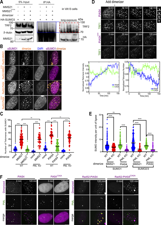
Recruiting SUMO ligases is sufficient for the formation of APBs and phase separation at telomeres. (A) Western blots for VA13 cells expressing Halo-GFP-TRF1, HA-SUMO3, and mCherry-eDHFR-MMS21/MMS21C215A with adding dimerizers for 5 h. SUMOylated proteins were captured with protein A/G beads following HA antibody incubation. The blue box indicates the zoom region. The red box indicates TRF2 bands being SUMOylated. (B and C) Representative images and quantification of SUMO1 localization at telomeres in U2OS WT and PML KO cells after dimerizing MMS21/MMS21C215A/PIAS4/PIAS4C342A/Rad52-MMS21/Rad52-MMS21C215A/Rad52-MMS21/Rad52-PIAS4C342A to telomeres for 5 h. Each dot represents one cell, three independent experiments, >54 cells per group. (D) Representative images (top) and quantification for telomere intensity (bottom left) and number (bottom right) in U2OS cells after dimerizing mCh-eDHFR-PIAS4 or PIAS4C342A to 3xHalo-GFP-TRF1 after the first time point. The zoomed-in images on the top right show the fusion of TRF1 foci after dimerizing PIAS4 (time interval 5 min). >19 cells per group, three independent experiments. (E) Quantification of SUMO1/2/3 intensity per unit of MMS21 or PIAS4 intensity on telomeres in U2OS WT and PML KO cells. Each dot represents one cell, three independent experiments, >52 cells per group. (F) Representative images of PML-Clover U2OS cells, expressing mCh-eDHFR-PIAS4/PIAS4C342A (left) or mCh-eDHFR-Rad52-PIAS4/PIAS4C342A (right) and 3xHalo-TRF1, with the addition of the dimerizer at the indicated time points. Yellow arrows indicate APBs. Scale bars, 5 μm or 1 μm for zoomed-in images. Source data are available for this figure: SourceData FS4.

Recruiting SUMO ligases is sufficient for the formation of APBs and phase separation at telomeres. (A) Western blots for VA13 cells expressing Halo-GFP-TRF1, HA-SUMO3, and mCherry-eDHFR-MMS21/MMS21C215A with adding dimerizers for 5 h. SUMOylated proteins were captured with protein A/G beads following HA antibody incubation. The blue box indicates the zoom region. The red box indicates TRF2 bands being SUMOylated. (B and C) Representative images and quantification of SUMO1 localization at telomeres in U2OS WT and PML KO cells after dimerizing MMS21/MMS21C215A/PIAS4/PIAS4C342A/Rad52-MMS21/Rad52-MMS21C215A/Rad52-MMS21/Rad52-PIAS4C342A to telomeres for 5 h. Each dot represents one cell, three independent experiments, >54 cells per group. (D) Representative images (top) and quantification for telomere intensity (bottom left) and number (bottom right) in U2OS cells after dimerizing mCh-eDHFR-PIAS4 or PIAS4C342A to 3xHalo-GFP-TRF1 after the first time point. The zoomed-in images on the top right show the fusion of TRF1 foci after dimerizing PIAS4 (time interval 5 min). >19 cells per group, three independent experiments. (E) Quantification of SUMO1/2/3 intensity per unit of MMS21 or PIAS4 intensity on telomeres in U2OS WT and PML KO cells. Each dot represents one cell, three independent experiments, >52 cells per group. (F) Representative images of PML-Clover U2OS cells, expressing mCh-eDHFR-PIAS4/PIAS4C342A (left) or mCh-eDHFR-Rad52-PIAS4/PIAS4C342A (right) and 3xHalo-TRF1, with the addition of the dimerizer at the indicated time points. Yellow arrows indicate APBs. Scale bars, 5 μm or 1 μm for zoomed-in images. Source data are available for this figure: SourceData FS4.
Telomere clustering after dimerizing MMS21 and PIAS4, but not after dimerizing MMS21C215A or PIAS4C342A, was seen with FISH (Fig. 6 D). Live imaging showed that both systems induced signatures of phase separation, including fusion events (Fig. 6 E left, Fig. S4 D top, Videos 5 and 6), increased telomere intensity (Fig. 6 E middle, Fig. S4 D bottom left), and decreased telomere number (Fig. 6 E right, Fig. S4 D bottom right), phenotypes similar to those that resulted from tethering SUMO to telomeres shown in our previous study (Zhao et al., 2024). Again, this effect was not seen with MMS21C215A or PIAS4C342A mutants (Fig. 6 E; Fig. S4 D; and Videos 7 and 8), confirming the necessity of SUMOylation for phase separation.
Dimerizing MMS21 to telomeres in U2OS cells. Movie for Fig. S4 D. Left: Composite of MMS21 (magenta) and TRF1 (green); middle: mCh-eDHFR-MMS21; right: 3xHalo-GFP-TRF1. The dimerizer was added to cells after the first time point to induce dimerization. The box shows a telomere fusion event. Scale bars, 5 μm.
Dimerizing MMS21 to telomeres in U2OS cells. Movie for Fig. S4 D. Left: Composite of MMS21 (magenta) and TRF1 (green); middle: mCh-eDHFR-MMS21; right: 3xHalo-GFP-TRF1. The dimerizer was added to cells after the first time point to induce dimerization. The box shows a telomere fusion event. Scale bars, 5 μm.
Dimerizing MMS21(C215A) to telomeres in U2OS cells. Movie for Fig. S4 D. Left: Composite of MMS21(C215A) (magenta) and TRF1 (green); middle: mCh-eDHFR-MMS21(C215A); right: 3xHalo-GFP-TRF1. The dimerizer was added to cells after the first time point to induce dimerization. Scale bars, 5 μm.
Dimerizing MMS21(C215A) to telomeres in U2OS cells. Movie for Fig. S4 D. Left: Composite of MMS21(C215A) (magenta) and TRF1 (green); middle: mCh-eDHFR-MMS21(C215A); right: 3xHalo-GFP-TRF1. The dimerizer was added to cells after the first time point to induce dimerization. Scale bars, 5 μm.
Dimerizing PIAS4 to telomeres in U2OS cells. Movie for Fig. S4 D. Left: Composite of PIAS4 (magenta) and TRF1 (green); middle: mCh-eDHFR-PIAS4; right: 3xHalo-GFP-TRF1. The dimerizer was added to cells after the first time point to induce dimerization. The box shows a telomere fusion event. Scale bars, 5 μm.
Dimerizing PIAS4 to telomeres in U2OS cells. Movie for Fig. S4 D. Left: Composite of PIAS4 (magenta) and TRF1 (green); middle: mCh-eDHFR-PIAS4; right: 3xHalo-GFP-TRF1. The dimerizer was added to cells after the first time point to induce dimerization. The box shows a telomere fusion event. Scale bars, 5 μm.
Dimerizing PIAS4(C342A) to telomeres in U2OS cells. Movie for Fig. S4 D. Left: Composite of PIAS4(C342A) (magenta) and TRF1 (green); middle: mCh-eDHFR-PIAS4(C342A); right: 3xHalo-GFP-TRF1. The dimerizer was added to cells after the first time point to induce dimerization. Scale bars, 5 μm.
Dimerizing PIAS4(C342A) to telomeres in U2OS cells. Movie for Fig. S4 D. Left: Composite of PIAS4(C342A) (magenta) and TRF1 (green); middle: mCh-eDHFR-PIAS4(C342A); right: 3xHalo-GFP-TRF1. The dimerizer was added to cells after the first time point to induce dimerization. Scale bars, 5 μm.
Similar observations were made in PML KO U2OS cells (Fig. 6, B and C), though SUMO intensity per unit of ligase recruited to telomeres (Fig. S4 E) and the degree of telomere clustering were less compared with those seen in PML-containing cells (Fig. 6 D). This result indicates that PML helps to enrich SUMO on telomeres to promote telomere clustering.
Recruiting SUMO ligases to telomeres forms APBs mainly via fusion
Recruiting MMS21 and PIAS4 to telomeres in U2OS cells formed more APBs in U2OS, SaoS2, and VA13 cells, whereas recruiting MMS21C215A or PIAS4C342A did not (Fig. 7, A and B). In all assays, the two SUMO ligases produced comparable results (Fig. 6, C and D; and Fig. 7 B), indicating that SUMOylation at telomeres rather than ligase identity is important for APB formation and other ALT features.

Recruiting SUMO ligases fused to Rad52 enhances APB nucleation. (A and B) Representative images and quantification of PML localization at telomeres in U2OS, SaoS2, and VA13 cells after dimerizing MMS21/MMS21C215A, PIAS4/PIAS4C342A, Rad52-MMS21/PIAS4, or Rad52-MMS21C215A/PIAS4C342A for 5 h. Each dot represents one cell, three independent experiments, >55 cells per group. (C) Representative images of PML-Clover U2OS cells expressing mCh-eDHFR-MMS21/MMS21C215A (left) or Rad52-MMS21/MMS21C215A (right) and 3xHalo-TRF1 with the addition of the dimerizer at indicated time points. Yellow arrows indicate APBs. Scale bars, 5 μm. (D) Quantification of newly formed APBs per cell after dimerizing MMS21/MMS21C215A/Rad52-MMS21/Rad52- MMS21C215A/PIAS4/PIAS4C342A/Rad52-PIAS4/Rad52-PIAS4C342A to telomeres in PML-Clover U2OS cells for 2 h. More than 25 cells per group, three independent experiments. (E) Western blot for U2OS cells expressing Halo-GFP-TRF1, HA-SUMO3, mCherry-eDHFR-MMS21, or Rad52-MM21, after adding the dimerizer for 5-h. SUMOylated proteins were captured with protein A/G beads following HA antibody incubation. The red box indicates SUMOylated TRF2. (F) Quantification of SUMO1/2/3 intensity per unit of MMS21/PIAS4 or Rad52-MMS21/PIAS4/MMS21C215A/PIAS4C342A on telomeres after dimerizing for 5 h in U2OS cells. Each dot represents one cell, three independent experiments, >47 cells per group. (G) ChIP-qRCR assay detecting PML binding to telomere DNA from indicated chromatin in U2OS cells with no dimerization, dimerizing MMS21, or Rad52-MMS21 to telomeres for 5 h, three independent experiments. (H) Quantification of telomere numbers in U2OS cells after dimerizing MMS21/PIAS4/Rad52-MMS21/Rad52-MMS21C215A/Rad52-PIAS4/Rad52-PIAS4C342A to telomeres for 5 h. Each dot represents one cell, three independent experiments, >40 cells per group. (I) APB formation time for nucleation and fusion after dimerizing the indicated proteins for 2 h in U2OS PML-Clover cells. Each dot represents one cell, three independent experiments, >36 cells per group. Source data are available for this figure: SourceData F7.
Recruiting SUMO ligases fused to Rad52 enhances APB nucleation. (A and B) Representative images and quantification of PML localization at telomeres in U2OS, SaoS2, and VA13 cells after dimerizing MMS21/MMS21C215A, PIAS4/PIAS4C342A, Rad52-MMS21/PIAS4, or Rad52-MMS21C215A/PIAS4C342A for 5 h. Each dot represents one cell, three independent experiments, >55 cells per group. (C) Representative images of PML-Clover U2OS cells expressing mCh-eDHFR-MMS21/MMS21C215A (left) or Rad52-MMS21/MMS21C215A (right) and 3xHalo-TRF1 with the addition of the dimerizer at indicated time points. Yellow arrows indicate APBs. Scale bars, 5 μm. (D) Quantification of newly formed APBs per cell after dimerizing MMS21/MMS21C215A/Rad52-MMS21/Rad52- MMS21C215A/PIAS4/PIAS4C342A/Rad52-PIAS4/Rad52-PIAS4C342A to telomeres in PML-Clover U2OS cells for 2 h. More than 25 cells per group, three independent experiments. (E) Western blot for U2OS cells expressing Halo-GFP-TRF1, HA-SUMO3, mCherry-eDHFR-MMS21, or Rad52-MM21, after adding the dimerizer for 5-h. SUMOylated proteins were captured with protein A/G beads following HA antibody incubation. The red box indicates SUMOylated TRF2. (F) Quantification of SUMO1/2/3 intensity per unit of MMS21/PIAS4 or Rad52-MMS21/PIAS4/MMS21C215A/PIAS4C342A on telomeres after dimerizing for 5 h in U2OS cells. Each dot represents one cell, three independent experiments, >47 cells per group. (G) ChIP-qRCR assay detecting PML binding to telomere DNA from indicated chromatin in U2OS cells with no dimerization, dimerizing MMS21, or Rad52-MMS21 to telomeres for 5 h, three independent experiments. (H) Quantification of telomere numbers in U2OS cells after dimerizing MMS21/PIAS4/Rad52-MMS21/Rad52-MMS21C215A/Rad52-PIAS4/Rad52-PIAS4C342A to telomeres for 5 h. Each dot represents one cell, three independent experiments, >40 cells per group. (I) APB formation time for nucleation and fusion after dimerizing the indicated proteins for 2 h in U2OS PML-Clover cells. Each dot represents one cell, three independent experiments, >36 cells per group. Source data are available for this figure: SourceData F7.
Using PML-Clover–containing cells to follow APB formation after ligase tethering with live imaging, we found that MMS21 and PIAS4 targeting to telomeres led to APB formation both by fusion and nucleation, while MMS21C215A or PIAS4C342A had minimal effects (Fig. 7 C left, Fig. 7 D; Fig. S4 F left), confirming the observation from fixed imaging that SUMOylation is necessary for APB formation (Fig. 7 B). MMS21’s effects were slightly more potent than PIAS4 during the 2 h of live imaging (Fig. 7 D), which differs from the fixed imaging after 5 h of dimerization that showed a similar level of APB formation (Fig. 7 B). This difference may indicate that MMS21 is more efficient at promoting APB formation at short time scales, likely caused by differences in ligase activity and/or available substrates. Moreover, recruiting both ligases led to APB formation by fusion more than by nucleation (Fig. 7 D), similar to the outcomes upon TRF1–FokI induction. The total number of APBs formed was also similar to that induced by TRF1–FokI (Fig. 1 D), indicating that SUMO enrichment by ligase recruitment is comparable with telomere break induction. Together, these data show that the enrichment of the two SUMO ligases is equally sufficient to induce APB formation but with fusion more so than nucleation.
Recruiting Rad52 together with SUMO ligases increases APB nucleation
The combined results using different SUMO enrichment or SUMO depletion regimes are consistent among themselves and support the notion that telomeres with lower SUMO levels support APB fusion but that a higher SUMO level is required for APB nucleation. If this is true, one would predict that while APB nucleation is less prominent than APB fusion in the SUMO ligase telomere targeting regime, further increased telomere–SUMO levels could result in more APB nucleation. To test this prediction, we targeted ligases together with Rad52, a repair protein implicated in ALT that can increase ALT telomere SUMO levels, as shown previously (Zhao et al., 2024). We confirmed that TRF2 SUMOylation increased upon dual tethering of MMS21 and Rad52 (Rad52-MMS21) to telomeres compared with only MMS21 by co-expressing HA-SUMO3 and pulling down HA (Fig. 7 E). Consistently, SUMO1 and SUMO2/3 intensities per unit of MMS21 and PIAS4 recruited were higher when Rad52 was introduced compared with when just MMS21 and PIAS4 or their enzymatically dead variants, MMS21C215A and PIAS4C342A, were targeted to telomeres (Fig. 7 F).
Significantly, dual targeting Rad52 and ligases to telomeres was associated with more APB formation in all three ALT cell lines, U2OS, SaoS2, and VA13 (Fig. 7, A and B). We also performed chromatin immunoprecipitation (ChIP) assay to assess telomere-associated PML protein on 10q, 13q, and 15q. We found that Rad52-MMS21 tethering enriched more PML on these telomeres than MMS21 tethering (Fig. 7 G), agreeing with results from the imaging data (Fig. 7 B). However, regarding the degree of telomere clustering, the dual targeting system has a comparable effect as ligase targeting alone (Fig. 7 H), likely because the SUMO level is no longer important for telomere clustering once the threshold for phase separation, which is required for telomere clustering, is reached. Finally, compared with MMS21 or PIAS4 alone, dual targeting of Rad52 with ligases led to more and faster APB nucleation than recruiting MMS21 or PIAS4 alone (Fig. 7, C, D, and I; and Fig S4 F). This result supports the prediction from our model that higher SUMO levels favor APB formation via nucleation.
APB nucleation is associated with stronger BLM recruitment to telomeres and telomeric DNA synthesis
Finally, we address possible functional consequences of the two APB formation pathways. Given the established role of APBs in recruiting BLM to ALT telomeres (Loe et al., 2020), we examine BLM enrichment on telomeres in different situations. As shown above, telomere recruitment of SUMO3, Rad52-MMS21, and Rad52-PIAS4 mainly leads to APB nucleation. For simplicity, we will refer to these tethering strategies as APB nucleation regimes. On the other hand, recruiting SIM, MMS21, and PIAS4 to telomeres mainly results in APB fusion and, thus, is collectively referred to as APB fusion regimes. IF showed that APB nucleation regimes led to more BLM on telomeres than the APB fusion regimes (Fig. 8, A and B). This was confirmed using an APEX proximity labeling assay with an APEX-Halo-TRF1 stable U2OS cell line to examine BLM associated with biotinylated TRF1 (Fig. 8 C). In addition, APB nucleation regimes led to more telomeric DNA synthesis than the APB fusion regimes (Fig. 8, A and D; and Fig. S5 A). Collectively, these results indicate that APB nucleation is more efficient at localizing BLM to telomeres for ALT telomeric DNA synthesis.

APB nucleation localizes BLM to telomeres more efficiently. (A) Representative images of EdU and BLM staining on telomeres after dimerizing SUMO3 (WT)/SIM/MMS21/Rad52-MMS21/Rad52-MMS21C215A in U2OS cells for 5 h. Yellow errors indicate EdU signal on telomeres or BLM localization on telomeres. Scale bars, 5 μm. (B) Quantification of endogenous BLM localization on telomeres after dimerizing SUMO3 (WT)/SIM/MMS21/MMS21C215A/Rad52-MMS21/Rad52-MMS21C215A/PIAS4/PIAS4C342A/Rad52-PIAS4/Rad52-PIAS4C342A in U2OS, SaoS2, and VA13 cells for 5 h. Each dot represents one independent experiment, >24 cells per group, three independent experiments. (C) IP for BLM on telomeres in APEX-Halo-TRF1 U2OS cells dimerized with mCherry-eDHFR-SIM/MMS21/SUMO3 (WT)/Rad52-MMS21. Biotinylated telomere proteins were captured with streptavidin beads. Cells were immunoblotted with anti-BLM, mCherry, and β-actin. The red box indicates SUMOylated BLM. (D) Quantification of EdU staining on telomeres after dimerizing SUMO3 (WT)/SIM/MMS21/PIAS4/Rad52-MMS21/Rad52-MMS21C215A/Rad52-PIAS4/Rad52-PIAS4C342A in U2OS, SaoS2, and VA13 cells for 5 h. Each dot represents one cell, three independent experiments, >51 cells per group. (E and F) Representative images and quantification of EdU on telomeres after dimerizing SUMO3 (WT)/SIM/MMS21/PIAS4 and their indicated mutants for 5 h in U2OS cells, with/without GFP-BLM overexpression. Yellow arrows indicate EdU localization at telomeres. Each dot represents one cell, three independent experiments, >47 cells per group. Scale bars, 5 μm. Source data are available for this figure: SourceData F8.
APB nucleation localizes BLM to telomeres more efficiently. (A) Representative images of EdU and BLM staining on telomeres after dimerizing SUMO3 (WT)/SIM/MMS21/Rad52-MMS21/Rad52-MMS21C215A in U2OS cells for 5 h. Yellow errors indicate EdU signal on telomeres or BLM localization on telomeres. Scale bars, 5 μm. (B) Quantification of endogenous BLM localization on telomeres after dimerizing SUMO3 (WT)/SIM/MMS21/MMS21C215A/Rad52-MMS21/Rad52-MMS21C215A/PIAS4/PIAS4C342A/Rad52-PIAS4/Rad52-PIAS4C342A in U2OS, SaoS2, and VA13 cells for 5 h. Each dot represents one independent experiment, >24 cells per group, three independent experiments. (C) IP for BLM on telomeres in APEX-Halo-TRF1 U2OS cells dimerized with mCherry-eDHFR-SIM/MMS21/SUMO3 (WT)/Rad52-MMS21. Biotinylated telomere proteins were captured with streptavidin beads. Cells were immunoblotted with anti-BLM, mCherry, and β-actin. The red box indicates SUMOylated BLM. (D) Quantification of EdU staining on telomeres after dimerizing SUMO3 (WT)/SIM/MMS21/PIAS4/Rad52-MMS21/Rad52-MMS21C215A/Rad52-PIAS4/Rad52-PIAS4C342A in U2OS, SaoS2, and VA13 cells for 5 h. Each dot represents one cell, three independent experiments, >51 cells per group. (E and F) Representative images and quantification of EdU on telomeres after dimerizing SUMO3 (WT)/SIM/MMS21/PIAS4 and their indicated mutants for 5 h in U2OS cells, with/without GFP-BLM overexpression. Yellow arrows indicate EdU localization at telomeres. Each dot represents one cell, three independent experiments, >47 cells per group. Scale bars, 5 μm. Source data are available for this figure: SourceData F8.

BLM overexpression is sufficient for telomeric DNA synthesis due to the lack of APB nucleation. (A) Representative images of EdU and BLM on telomeres after dimerizing PIAS4/Rad52-PIAS4/Rad52-PIAS4C342A in U2OS cells for 5 h. (B–D) Representative images and quantifications of BLM localization and EdU on telomeres after expressing BLM WT, BLMTKR, BLMSIM, and BLMTKR-SIM in U2OS BLM KO cells. (E) Representative images of BLM localization at telomeres after adding dimerizers at the indicated time points in U2OS cells expressing mCh-eDHFR-SIM or SUMO3 (WT), 3xHalo-TRF1, and GFP-BLM. Yellow arrows indicate BLM localization at telomeres. (F) Quantification of GFP-BLM localization on telomeres after dimerizing SUMO3 (WT)/SIM/MMS21/MMS21C215A/PIAS4/PIAS4C342A to telomeres for 5 h in U2OS cells expressing GFP-BLM. Each dot represents one independent experiment, >28 cells per group, three independent experiments. (G) Representative images of BLM localization at telomeres after adding dimerizers at the indicated time points in U2OS cells expressing mCh-eDHFR-MMS21/MMS21C215A or PIAS4/PIAS4C342A, 3xHalo-TRF1, and GFP-BLM. (H) Representative images of EdU staining on telomeres after dimerizing SUMO3FAAA/SIMVADA/MMS21C215A/PIAS4C342A for 5 h in U2OS cells expressing GFP-BLM. Scale bars, 5 μm.

BLM overexpression is sufficient for telomeric DNA synthesis due to the lack of APB nucleation. (A) Representative images of EdU and BLM on telomeres after dimerizing PIAS4/Rad52-PIAS4/Rad52-PIAS4C342A in U2OS cells for 5 h. (B–D) Representative images and quantifications of BLM localization and EdU on telomeres after expressing BLM WT, BLMTKR, BLMSIM, and BLMTKR-SIM in U2OS BLM KO cells. (E) Representative images of BLM localization at telomeres after adding dimerizers at the indicated time points in U2OS cells expressing mCh-eDHFR-SIM or SUMO3 (WT), 3xHalo-TRF1, and GFP-BLM. Yellow arrows indicate BLM localization at telomeres. (F) Quantification of GFP-BLM localization on telomeres after dimerizing SUMO3 (WT)/SIM/MMS21/MMS21C215A/PIAS4/PIAS4C342A to telomeres for 5 h in U2OS cells expressing GFP-BLM. Each dot represents one independent experiment, >28 cells per group, three independent experiments. (G) Representative images of BLM localization at telomeres after adding dimerizers at the indicated time points in U2OS cells expressing mCh-eDHFR-MMS21/MMS21C215A or PIAS4/PIAS4C342A, 3xHalo-TRF1, and GFP-BLM. (H) Representative images of EdU staining on telomeres after dimerizing SUMO3FAAA/SIMVADA/MMS21C215A/PIAS4C342A for 5 h in U2OS cells expressing GFP-BLM. Scale bars, 5 μm.
Next, we determined whether BLM recruitment to telomeres depends on its SUMOylation sites and SIM. We expressed BLM WT, its SUMOylation-defective mutant (triple lysine K to arginine R: K317R, K331R, and K344R; BLMTKR), its SIM-defective mutant (QIDL to QADA, VICI to AACI; BLMSIM), and its SUMO- and SIM-defective mutant (BLMTKR-SIM) in BLM KO U2OS cells and dimerized SUMO3 or SIM to the telomeres (Min et al., 2019). We found that the BLMTKR and BLMSIM both decreased BLM localization on telomeres compared with WT for SUMO3 and SIM tethering, while the double mutants abolished its localization (Fig. S5, B and C). BLM single mutants also led to fewer EdU signals (Fig. S5, B and D). However, the double mutants (BLMTKR-SIM) did not show further reductions, likely due to lower sensitivity in EdU detection than in BLM telomere localization. These data suggest that the SUMOylation sites and SIM in BLM both contribute to its telomeric localization and its role in promoting telomere DNA synthesis.
To examine whether the key outcome of the APB nucleation regime is to enrich BLM on telomeres, we asked whether overexpressing BLM could rescue the lower level of telomeric DNA synthesis in the APB fusion regimes. This was indeed the case, as we found that more telomeres were enriched with BLM (Fig. S5, E–G) and had more robust telomeric DNA synthesis in the APB fusion regimes, such as recruiting MMS21, PIAS4, and SIM, after BLM overexpression (Fig. 8, E and F; and Fig. S5 H). Importantly, these effects were not seen after recruiting MMS21C215A, PIAS4C342A, SUMO3FAAA, and SIMVADA, supporting that both SUMO–SIM interaction and SUMOylation are required for BLM-mediated telomeric DNA synthesis. Interestingly, BLM overexpression after recruiting MMS21 and PIAS4 resulted in both a similar increase in BLM localization on telomeres (Fig. S5 F) and DNA synthesis (Fig. 8 F) as recruiting SUMO3, indicating that overexpressing BLM is sufficient to compensate for the defects of BLM recruitment and telomeric DNA synthesis due to the lack of APB nucleation.
APB formation pathway affects other ALT features
Given that the APB formation pathway determines BLM localization and telomeric DNA synthesis, we investigated whether the choice of pathway also correlates with other ALT features. First, we examined C-circle levels, a unique ALT feature associated with ALT telomere DNA synthesis. We found that recruiting SUMO3 resulted in greater C-circle formation than recruiting SIM or inducing FokI on telomeres in U2OS cells (Fig. 9, A and B). Additionally, knocking down SUMO2/3 reduced C-circle levels in U2OS cells more than knocking down SUMO1 (Fig. 9, A and B), which aligns with previous findings showing that SUMO2/3 contributes more to the ALT pathway.

APB formation pathway affects other ALT features. (A and B) C-circle dot blot and quantification without treatment, inducing FokI, and dimerizing SUMO3 (WT)/SIM, or after transfecting siSUMO1 or siSUMO2/3 for 2 days in U2OS cells. (C) Representative images of TRF1, ssTelo-C staining after dimerizing SUMO3 (WT)/SIM in SaoS2 cells for 5 h. (D) Representative images of TRF1, ssTelo-C staining after transfecting siSUMO1 or siSUMO2/3 in VA13 cells for 2 days. Scale bars, 5 μm. (E) Quantification of ssTelo-C after dimerizing SUMO3 (WT), SIM, or knocking down SUMO1 and SUMO2/3 in U2OS, Saos2, and VA13 cells. Each dot represents one cell, three independent experiments, >31 cells per group. (F) Proposed model. Top: Telomeres with more DNA damage recruit more DNA repair factors as SUMO substrates, resulting in the enrichment of SUMO on telomeres by MMS21-mediated polySUMOylation, which then recruits PML protein to nucleate PML-NBs on telomeres to promote efficient BLM recruitment and telomere elongation. Bottom: Telomeres with less DNA damage recruit fewer DNA repair factors as SUMO substrates, resulting in low enrichment of SUMO on telomeres, which cannot recruit PML protein to nucleate PML-NBs but can fuse with PML-NBs to form APBs, resulting in less efficient BLM recruitment and telomere elongation. Created with https://BioRender.com. Source data are available for this figure: SourceData F9.
APB formation pathway affects other ALT features. (A and B) C-circle dot blot and quantification without treatment, inducing FokI, and dimerizing SUMO3 (WT)/SIM, or after transfecting siSUMO1 or siSUMO2/3 for 2 days in U2OS cells. (C) Representative images of TRF1, ssTelo-C staining after dimerizing SUMO3 (WT)/SIM in SaoS2 cells for 5 h. (D) Representative images of TRF1, ssTelo-C staining after transfecting siSUMO1 or siSUMO2/3 in VA13 cells for 2 days. Scale bars, 5 μm. (E) Quantification of ssTelo-C after dimerizing SUMO3 (WT), SIM, or knocking down SUMO1 and SUMO2/3 in U2OS, Saos2, and VA13 cells. Each dot represents one cell, three independent experiments, >31 cells per group. (F) Proposed model. Top: Telomeres with more DNA damage recruit more DNA repair factors as SUMO substrates, resulting in the enrichment of SUMO on telomeres by MMS21-mediated polySUMOylation, which then recruits PML protein to nucleate PML-NBs on telomeres to promote efficient BLM recruitment and telomere elongation. Bottom: Telomeres with less DNA damage recruit fewer DNA repair factors as SUMO substrates, resulting in low enrichment of SUMO on telomeres, which cannot recruit PML protein to nucleate PML-NBs but can fuse with PML-NBs to form APBs, resulting in less efficient BLM recruitment and telomere elongation. Created with https://BioRender.com. Source data are available for this figure: SourceData F9.
Next, we tested another ALT feature, the level of single-strand C-rich telomere repeat (ssTelo-C), using a native FISH assay after recruiting SUMO3 and SIM to telomeres in U2OS, SaoS2, and VA13 cells. We found that recruiting SUMO3 produced more ssTelo-C than recruiting SIM in all three ALT cell lines (Fig. 9, C and E). In addition, knocking down SUMO2/3 resulted in a greater reduction of ssTelo-C than knocking down SUMO1 in the SaoS2 and VA13 cell lines (Fig. 9, D and E). Together, these data suggest that SUMO levels on the telomeres may influence several ALT features by impacting the APB formation pathway.
Discussion
Previous studies have revealed the requirement of SUMOylation in APB formation through modifying the telomere-binding proteins, DNA repair factors, and the PML protein (Brouwer et al., 2009; Chung et al., 2011; Potts and Yu, 2007; Zhang et al., 2020). In this study, we followed APB formation dynamics by initiating chemically inducible DNA damage or SUMO tethering to telomeres. Through time-lapse imaging, we reveal two pathways for APB formation: de novo nucleation of PML-NBs on telomeres and fusion of telomeres with existing PML-NBs (Fig. 9 F). Through mutagenesis experiments, we show that both pathways require SUMO–SIM interactions at both the telomere and on the PML protein, highlighting the importance of SUMO–SIM interactions in APB formation.
We also show that both APB formation pathways can occur in the same cell, indicating that the determinant for pathway choice is at the telomere level. Through tethering experiments, we show that the identities of SUMO paralogs and SUMO ligases do not affect APB formation pathway choice. Instead, the SUMO level of telomeres matters: telomeres with high SUMO levels favor APB formation via nucleation, while telomeres with low SUMO levels form APBs via fusion. This is likely because greater SUMO levels on telomeres are needed to compete with the existing PML-NBs for the free PML protein in the nucleoplasm for de novo nucleation of PML-NBs on telomeres. Supporting the importance of SUMO level, knocking down the polySUMO chain forming SUMO2/3 reduces APB formation via nucleation more than the non-polySUMO chain forming SUMO1. In addition, tethering DNA repair factor Rad52 with SUMO ligases can increase SUMO levels and promote APB formation via nucleation, indicating that the amount of DNA repair factors—likely caused by different degrees of DNA damage on each telomere—can contribute to the pathway choice of APB formation (Fig. 9 F). These results agree with the previously reported promiscuous nature of SUMOylation, where SUMO ligases modify a group of substrates in the vicinity, and it is this group modification, not the modification of individual proteins, that is functionally important (Dhingra and Zhao, 2019; Hu and Parvin, 2014; Psakhye and Jentsch, 2012; Jentsch and Psakhye, 2013).
We further demonstrate that the functional relevance of the APB formation pathway lies in how it contributes to BLM recruitment to telomeres, one of the key roles of APBs in ALT (Loe et al., 2020). Our data show that both pathways of APB formation can help recruit BLM to telomeres, but APB formation via nucleation has a stronger effect (Fig. 9 F). This is likely because telomeres with SUMO levels high enough to compete with the existing PML-NBs for free PML protein are also likely to form bigger PML-NBs, which in turn recruit more BLM. As a result of more BLM recruitment, telomeres that can form APBs via nucleation have more robust telomeric DNA synthesis and other ALT features, such as telomere clustering. However, it is possible that APB formation contributes to some of these features through mechanisms independent of BLM recruitment. For example, the APB formation pathway might influence telomere mobility differently, thereby affecting telomere clustering, which could be verified by tracking telomere mobility in future studies. Nevertheless, our results indicate that telomeres with APBs formed via nucleation are likely to elongate more than telomeres with APBs formed via fusion in the same cell, leading to heterogeneity in telomere length that is often observed in ALT cells. In supporting this prediction, ALT cells that lack APBs are shown to lose telomere length heterogeneity (Loe et al., 2020).
We also show that artificially elevated SUMO, but not DNA damage, can induce PML-NB formation at centromeres. These findings may explain why PML-NBs are associated with ALT telomeres more often than non-telomere DNA damage sites (Chang et al., 2018; Dhingra and Zhao, 2019; Hu and Parvin, 2014; Psakhye and Jentsch, 2012; Yeung et al., 2012). First, ALT telomeres are known to experience high replication stress that can result in DNA damage (Lu and Pickett, 2022). This can lead to greater enrichment of repair proteins that can be SUMOylated at telomeres. In addition, ALT cells have some extremely long telomeres (Bryan et al., 1995). These long telomeres harbor more telomere-binding proteins that can be promiscuously SUMOylated in response to damage-induced SUMO ligase localization to telomeres. Through either SUMOylation of repair proteins or telomere-binding proteins, the enhanced SUMO accumulation can promote PML-NB nucleation on telomeres. On the other hand, telomeres are relatively mobile compared with other regions of the genome, which offers a high chance for the SUMO-coated telomeres to collide with PML-NBs. In supporting this kinetic model for PML-NB–damage site association, PML-NBs are not found at easy-to-repair DNA or early stage of damage sites, but a stable association of PML-NBs with damage foci can be observed at difficult-to-repair damage sites (Rodier et al., 2011; Vancurova et al., 2019; Hornofova et al., 2023; Kleijwegt et al., 2023). Future efforts to compare PML-NB formation dynamics at damaged genomic loci beyond centromeres with ALT telomeres can help test these predictions.
In summary, our work fills several gaps in our knowledge regarding APB biogenesis in ALT. The results may also conceptually impact models of the interplay between SUMO and PML-NBs in other pathways, such as DNA replication (Stubbe et al., 2020) and transcription (Pavan Kumar et al., 2007; Tan et al., 2008; Gasser and Stutz. 2023; Salsman et al., 2017), where both SUMO and PML-NBs are known to play regulatory roles. In addition, our discoveries on the mechanisms of PML-NB localization to ALT telomeres may contribute to the understanding of other condensate–chromatin associations, such as the dynamic contacts between nuclear speckles and transcriptional sites (Zhong et al., 2000).
Materials and methods
Cell culture
All experiments were performed with U2OS cells unless otherwise stated. PML KO and BLM KO U2OS cells were gifts from Dr. Eros Lazzerini Denchi. VA13 and SaoS2 cells were gifts from Dr. Roderick O’Sullivan. SUMO1 KO, SUMO2 KO, and SUMO2 KO+SUMO2 cells were from Dr. Michael J Matunis. U2OS cells with endogenous PML tagged with Clover (U2OS Clover-PML) were previously described (Pinder et al., 2015). Cells were cultured in growth medium (Dulbecco’s modified Eagle’s medium with 10% FBS and 1% penicillin–streptomycin) at 37°C in a humidified atmosphere with 5% CO2.
Plasmids
The plasmid for inducing DNA damage at telomeres (mCherry-TRF1–FokI) was previously published (Cho et al., 2014). 3xHalo-GFP-TRF1 was previously published (Zhang et al., 2020). SUMO1/2/3 WT for mCherry-eDHFR-SUMO came from plasmids gifted by Karsten Rippe (Chung et al., 2011). SIM and SUMO mutants for mCherry-eDHFR-SIM/SUMO were from plasmids gifted by Michael Rosen, where the SIM-interacting mutants of SUMO were generated by mutating the FKIK (SUMO3) or FKVK (SUMO1) to FAAA, nonconjugable SUMO modules were generated by mutating the C-terminal di-Gly motif to di-Val, and the mutant of SIM defective in SUMO binding was generated by mutating the VIDL sequence into VADA (Banani et al., 2016). The vector containing mCherry-eDHFR was from our published plasmid Mad1-mCherry-eDHFR (Zhang et al., 2017). PML WT and PML SUMOylation sites were from plasmids gifted by Michael Rosen (Banani et al., 2016). PML SIM mutant was kindly provided by Dr. Hung-Ying Kao and subcloned into the target backbone. GFP-BLM was from Addgene plasmid #80070. MMS21, PIAS4, mutants, CENPC, and LSD1 were introduced into target plasmids through in-fusion cloning (638948; Takara Bio). MMS21C215A and PIAS4C342A were generated with Q5 Site-Directed Mutagenesis Kit (E0554S; NEB). HA-SUMO3 plasmid was a gift from Dr. Roderick O’Sullivan. GFP BLM WT, TKR (SUMO-defective), SIM (SIM-defective), TKR/SIM (double mutant) were gifts from Dr. Jaewon Min. All the target plasmids in this study were derived from a plasmid that contains a CAG promoter for constitutive expression, obtained from E.V. Makeyev (Khandelia et al., 2011).
Synchronize cells to G2
Cells were first treated with 2 mM thymidine (T1895; Sigma-Aldrich) for 21 h, released into fresh medium for 4 h, and then treated with 15 μM CDK1i (RO-3306, cat# SML0569; Sigma-Aldrich) for 12 h.
siRNA transfection
siRNA for SUMO1 (sc-29498; Santa Cruz), SUMO2/3 (sc-37167; Santa Cruz), MMS21 (L-018070-00-0005; Horizon), and PIAS4 (L-006445-00-0005; Horizon) were purchased commercially. Cells were transfected with 100 nM of siRNA and RNAiMax (13778075; Thermo Fisher Scientific) diluted in OptiMEM (31-985-070; Life Technologies). The transfection medium was replaced with culture media 8 h later, and the transfection was repeated on day 2. Cells were collected and imaged at 48 h after the second-round transfection.
Protein dimerization and FokI induction
Design, synthesis, and storage of the dimerizer TFH were previously reported (Lackner et al., 2022). Dimerization to telomeres was performed as previously described (Zhao et al., 2021). Briefly, TFH was added directly to the growth medium to a final working concentration of 100 nM. Cells were incubated with the dimerizer-containing medium for the indicated times, followed by IF or FISH. For live imaging with protein dimerization, the dimerizers were first diluted to 200 nM in growth medium and then added to cell chambers to the working concentration after first-round imaging.
To induce damage on telomeres in cells transfected with mCherry-ER-DD-TRF1–FokI, Shield-1 (Cheminpharma LLC) and 4-hydroxytamoxifen (Sigma-Aldrich) at 1 μM were added for 2 h to allow TRF1 to enter the nucleus after first-round imaging, as previously described (Cho et al., 2014). SUMO inhibitor ML-792 (HY-108702) used to inhibit SUMOylation was purchased from Selleckchem.
IF and telomere DNA FISH
IF and FISH were performed following a previously published protocol (Zhao et al., 2021). Cells were fixed in 4% formaldehyde for 10 min at room temperature, followed by permeabilization in 0.5% Triton X-100 for 10 min. Cells were incubated with primary antibody at 4°C in a humidified chamber overnight and then with secondary antibody for 1 h at room temperature before washing and mounting. Primary antibodies were anti-TRF1 (1:100 dilution, Ab10579; Abcam), anti-CENPC (1:100 dilution, PA566154; Thermo Fisher Scientific), anti-SUMO1 (1:200 dilution, Ab32058; Abcam), anti-SUMO2/3 (1:200 dilution, Asm23; Cytoskeleton), anti-PML (1:100 dilution, sc966; Santa Cruz), anti-BLM (1:100 dilution, A300-110A; Bethyl), anti-PCNA (2586S, 1:1,000 dilution; Cell signaling), anti-γH2AX (1:1,000 dilution, 05-636; Millipore), and anti-RPA (1:100 dilution, NB600-565; Novus). For IF-FISH, coverslips were first stained with primary and secondary antibodies, then fixed again in 4% formaldehyde for 10 min at room temperature. Coverslips were then dehydrated in an ethanol series (70%, 80%, and 90%, 2 min each) and incubated with a TelC-Alexa488 or TelC-Cy3 PNA probe (F1004, F1002; PNA Bio) at 75°C for 5 min and then overnight in a humidified chamber at room temperature. Coverslips were then washed and mounted for imaging.
Telomeric DNA synthesis detection by EdU
Following transfection, cells were pulsed with EdU (10 μM) along with protein dimerization or DNA damage induction for 6 h before harvest. Cells on glass coverslips were washed twice in PBS and fixed with 4% PFA for 10 min. Cells were permeabilized with 0.3% (vol/vol) Triton X-100 for 5 min. The Click-IT Plus EdU Cell Proliferation Kit with Alexa Fluor 647 (C10635; Invitrogen) was applied to cells for 30 min to detect EdU.
ssTelo-C staining
Cells were grown on coverslips and fixed in 4% PFA for 10 min. Coverslips were then incubated with 500 µg/ml RNase A in blocking solution (1 mg/ml BSA, 3% goat serum, 0.1% Triton X-100, and 1 mM EDTA in PBS) for 1 h at 37°C. Coverslips were dehydrated in an ethanol series (70%, 90%, and 100%) and hybridized at room temperature with a TelG-Alexa488 PNA probe (F1008; PNA Bio) in hybridization buffer (70% formamide, 1 mg/ml blocking reagent [11096176001; Roche], and 10 mM Tris-HCl at pH 7.2). After washing, coverslips were stained with DAPI and imaged. A minimum of three independent experiments was performed for each datum shown unless indicated otherwise. For each experiment, at least 30 nuclei were imaged.
Cell imaging and image processing
Imaging acquisition was performed as previously described (Xu et al., 2024). For live imaging, cells were seeded on 22 × 22-mm glass coverslips coated with poly-D-lysine (P1024; Sigma-Aldrich). When ready for imaging, coverslips were mounted in magnetic chambers (Chamlide CM-S22-1; LCI) with cells maintained in a normal medium supplemented with 10% FBS and 1% penicillin/streptomycin at 37°C on a heated stage in an environmental chamber (TOKAI HIT Co., Ltd.). Images were acquired with a microscope (ECLIPSE Ti2) with a 100 × 1.4 NA objective, a 16 XY Piezo-Z stage (Nikon Instruments, Inc.), a spinning disk (Yokogawa), an electron multiplier charge-coupled device camera (IXON-L-897), and a laser merge module that was equipped with 488, 561, 594, and 630 nm lasers controlled by NIS-Elements Advanced Research. For both the fixed cells and live imaging, images were taken with 0.5 μm spacing between Z slices, for a total of 8 μm. For movies, images were taken at 5-min intervals for up to 3 h.
Western blotting
Cells were harvested with trypsin, quickly washed in PBS, and directly lysed in 4X NuPage LDS sample buffer (NP0007; Thermo Fisher Scientific). The resulting whole-cell lysates were analyzed by western blotting with the following primary antibodies: SUMO1 (1:1,000 dilution, Ab32058; Abcam), SUMO2/3 (1:1,000 dilution, Asm23; Cytoskeleton), MMS21 (1:1,000 dilution, A304-129A; Bethyl), PIAS4 (1:1,000 dilution, 4392; Cell Signaling), TRF2 (1:1,000 dilution, NBP1-86911; Novus), mCherry (1:1,000 dilution, ab183628; Abcam), BLM (1:1,000 dilution, A300-110A; Bethyl), HA (1:1,000 dilution, 3724S; Cell signaling), and β-Actin (1:1,000 dilution, Ab8226; Abcam). The HRP signal was visualized with Super Signal ECL substrate (34095; Pierce) per the manufacturer’s instructions.
SUMOylation protein pull-down
Approximately 4 × 10⁶ cells were scraped and lysed with 300 μl NP40 buffer (FNN0021; Thermo Fisher Scientific) with protease inhibitor (78430; Thermo Fisher Scientific) and N-Ethylmaleimide (E3876; Sigma-Aldrich) to prevent SUMO deconjugation. A fraction (20 μl) of the cell suspensions was kept as input. 3 µl of HA antibody (3724S; Cell signaling) was added to each group and was rotated at 4°C for 2 h. Protein A/G beads (88802; Thermo Fisher Scientific) (12 μl for each sample) were washed three times with NP40 buffer and then were incubated together with the lysates and antibody at 4°C overnight. Subsequently, Protein A/G beads were washed three times. Finally, the proteins captured by Protein A/G beads were eluted by boiling in LDS Sample Buffer (NP0007; Invitrogen). The levels of indicated proteins in input and pull-down samples were analyzed by western blot.
Proximity biotinylation using the peroxidase APEX2
Biotin phenol (SML2135; Sigma-Aldrich) was dissolved in dimethyl sulfoxide as a 500 mM stock solution and was added directly to cell culture media to a final concentration of 500 μM. H2O2 (H1009; Sigma-Aldrich) was spiked into the cell culture media to a final concentration of 1 mM to induce biotinylation. After 1 min of very gentle swirling, the media was decanted as quickly as possible, and the cells were washed three times with PBS containing 100 mM sodium azide (S2002; Sigma-Aldrich), 100 mM sodium ascorbate (PHR1279; Sigma-Aldrich), and 50 mM 6-hydroxy-2,5,7,8-tetramethylchroman-2- carboxylic acid (AC218940010; Tocris). Approximately 4 × 10⁶ cells were scraped and lysed with 300 μl RIPA buffer (89900; Thermo Fisher Scientific) with protease inhibitor (78430; Thermo Fisher Scientific) and N-Ethylmaleimide (E3876; Sigma-Aldrich) to prevent SUMO deconjugation. A fraction (20 μl) of the cell suspensions was kept as input. Streptavidin C1 beads (65001; Invitrogen) (12 μl for each sample) were washed three times with RIPA denaturing buffer and were then incubated together with the lysates at 4°C overnight. Subsequently, Streptavidin C1 beads were washed three times. Finally, the proteins captured by Streptavidin C1 beads were eluted by boiling in LDS Sample Buffer (NP0007; Invitrogen). The levels of indicated proteins in input and pull-down samples were analyzed by western blot.
Quantitative real-time PCR
Total RNAs were isolated using the RNA miniprep (Zymo; NC1047980), and 1 μg of RNAs was used for reverse transcription with the iScript cDNA Synthesis kit (Cat# 1708891; Bio-Rad). Quantitative PCR reactions were performed with equal amounts of cDNA using the DyNAmo SYBR Green qPCR kit (Cat# F410; Thermo Fisher Scientific). Signals were detected by the QuantStudio 3 (A28567; Thermo Fisher Scientific). GAPDH served as the internal control. The sequences of individual primers for the selected genes are provided in Table S1.
ChIP assay
The ChIP assay was conducted as described using a ChIP-IT High Sensitivity kit (53040; Active Motif). Briefly, ∼4 × 10⁶ cells were fixed with 1% formaldehyde for 10 min and then quenched in glycine for an additional 5 min at room temperature. Cells were lysed in extraction buffer on ice to obtain a nuclear pellet, followed by incubation with micrococcal nuclease (1 μl, 20 min at 37 °C) to fragment genomic DNAs. To ensure complete lysis of the nucleus, further sonication was performed using three sets of 20-s pulses. The resulting sheared DNAs were subjected to immunoprecipitation using normal IgG and anti-PML (sc966; Santa Cruz). Quantitative real-time PCR analysis was conducted to assess the immunoprecipitated telomere DNAs. The sequences of individual primers for the selected genes are provided in Table S1.
C-circle assay
Genomic DNA was purified, digested with AluI and MboI, and cleaned using phenol-chloroform extraction and precipitation. DNA was diluted in ultraclean water, and concentrations were measured to the indicated amounts (30, 15, and 7.5 ng) using a NanoDrop (Thermo Fisher Scientific). Samples (10 μl) were combined with 10 μl of BSA (NEB; 0.2 mg ml–1); 01 % Tween; 0.2 mM each of dATP, dGTP, and dTTP; and 1× Φ29 Buffer (NEB) in the presence or absence of 7.5 U of ΦDNA polymerase (NEB). Samples were incubated at 30°C for 8 h and then at 65°C for 20 min. Reaction products were diluted to 100 μl with 2× SSC and dot blotted onto a nylon membrane soaked in 2× SSC. DNA was UV cross-linked onto the membrane using UV light and hybridized with a biotin-labeled (CCCTAA)4 oligonucleotide probe (F2001; PNA Bio) to detect C-circle amplification products. The HRP signal was visualized with the Chemiluminescent Nucleic Acid Detection Module Kit (Thermo Fisher Scientific) as per the manufacturer’s instructions.
Statistical methods
All error bars represent means ± SEM. Statistical analyses were performed using Prism 10.0 (GraphPad software). Two-tailed unpaired t tests have been used for all tests. Statistical significance: P > 0.05; *P < 0.05; **P < 0.01; ***P < 0.001.
Online supplemental material
Fig. S1 is associated with Figs. 1 and 3, which show that the mutants that disrupt SUMO–SIM interactions could not lead to APB formation. Fig. S2 is associated with Fig. 4, which shows that the knocking down SUMO2/3 results in more defects in ALT features than knocking down SUMO1. Fig. S3 is associated with Fig. 5, which shows that the SUMO localization decreases more when knocking down MMS21, compared with PIAS4 in PMLKO cells. Fig S4 is associated with Figs. 6 and 7, which show that the APB nucleation is affected by SUMO ligase and SUMO substrate Rad52. Fig. S5 is associated with Fig. 8, which shows that the overexpressing BLM is sufficient for telomere DNA synthesis in the APB fusion regime. Video 1 shows the inducing DNA damage at telomeres in PML-Clover cells. Videos 2, 3, and 4 show the recruiting of SUMO3/SUMO3FAAA/SIM to telomeres in PML-Clover cells. Videos 5, 6, 7, and 8 show the dimerizing of MMS21/C215A/PIAS4/C342A to telomeres in PML-Clover cells. 3xHalo-TRF1 was co-transfected. Table S1 lists the primers used in this study.
Data availability
All data needed to evaluate the conclusions in the paper are present in the paper and the Online supplemental material. Raw data are available from the corresponding author upon reasonable request.
Acknowledgments
We thank Dr. Roger A. Greenberg (Department of Cancer Biology, Penn Center for Genome Integrity, Basser Center for BRCA, Perelman School of Medicine, University of Pennsylvania, Philadelphia, PA, USA), Dr. Eros Lazzerini Denchi (Laboratory of Genome Integrity, National Cancer Institute, National Institutes of Health, Bethesda, MD, USA), Dr. Michael J Matunis (Department of Biochemistry and Molecular Biology, Johns Hopkins University Bloomberg School of Public Health, Baltimore, MD, USA), Dr. Michael Rosen (Department of Biophysics and Howard Hughes Medical Institute, University of Texas Southwestern Medical Center, Dallas, TX, USA), Dr. Karsten Rippe (German Cancer Research Center (DKFZ) Heidelberg, Division of Chromatin Networks, Heidelberg, Germany), Dr. Roderick J. O’Sullivan (Department of Pharmacology and Chemical Biology, UPMC Hillman Cancer Center, University of Pittsburgh, Pittsburgh, PA, USA), Dr. Jaewon Min (Institute for Cancer Genetics, Columbia University Vagelos College of Physicians and Surgeons, New York, NY, USA), Eugene V. Makeyev (Center for Developmental Biology, King’s College London, London, UK), and Dr. Hung-Ying Kao (Departments of Biochemistry, Case Western Reserve University School of Medicine, Cleveland, OH, USA) for kindly gifting plasmids and cell lines.
This work was supported by the United States National Institutes of Health Grant U01CA260851 to Huaiying Zhang, GM118510 to David M. Chenoweth, R35 GM145260 to Xiaolan Zhao, GM060980 to Michael J. Matunis, and a Project Grant from the Canadian Institutes of Health Research PJT-156017 to Graham Dellaire.
Author contributions: Rongwei Zhao: conceptualization, data curation, formal analysis, investigation, methodology, project administration, resources, validation, visualization, and writing—original draft, review, and editing. Xiaoyang Yu: formal analysis, investigation, methodology, validation, visualization, and writing—review and editing. Tafadzwa Chigumira: formal analysis and investigation. Meng Xu: investigation. Allison Wivagg: data curation. Rachel M. Lackner: methodology. Jayme Salsman: methodology and resources. Graham Dellaire: resources and writing—review and editing. Michael J. Matunis: resources and writing—review and editing. David M. Chenoweth: methodology, resources, supervision, validation, and writing—review and editing. Xiaolan Zhao: conceptualization, funding acquisition, project administration, visualization, and writing—original draft, review, and editing. Huaiying Zhang: conceptualization, funding acquisition, project administration, resources, supervision, visualization, and writing—original draft, review, and editing.

