1-20 of 81588
Follow your search
Access your saved searches in your account

Would you like to receive an alert when new items match your search?
Close Modal
Sort by
Journal Articles
Images
Zbtb32 expression is selectively upregulated in Ttex tumor-infiltrating CD8+ T cells. (A) Fold change in the expression level of different TFs in Tpex and Ttex OT-I TILs in E.G7. (B) Volcanic plot showing the differential expression of TFs between Tpex and Ttex OT-I TILs in E.G7. (C) FeaturePlots showing Cd8a and Zbtb32 expression profiles in B16 TILs. (D) The expression levels of Zbtb32 and Cd8a in different clusters of B16 TILs. (E) The tSNE plot of scRNA-seq dataset, indicating global transcriptomic similarities of CD8+ TILs in B16 TME. (F) FeaturePlots showing expression profiles of Zbtb32 and other classic genes in B16 CD8+ TILs. (G)Zbtb32 and other classic genes expression levels in different clusters of B16 CD8+ TILs. (H) RT-qPCR analysis of Zbtb32 expression in Ly108+ and Tim-3+ CD8+ TILs (n = 3 for each group). GEO accessions: (A and B) GSE182035, (C–G) GSE122675. Unpaired two-tailed Student’s t test (H), ***P < 0.001. Data shown are a representative of at least two independent experiments.
Published: 06 February 2026
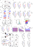
Figure 1. Zbtb32 expression is selectively upregulated in T tex tumor-infiltrating CD8 + T cells. (A) Fold change in the expression level of different TFs in Tpex and Ttex OT-I TILs in E.G7. (B) Volcanic plot showing the differential More about this image found in Zbtb32 expression is selectively upregulated in T ...
Images
The CD28-PI3K axis induces Zbtb32 expression after CD8+T cell activation. (A) The level of Zbtb32 expression was measured by RT-qPCR in activated CD8+ T cells in vitro (n = 3 for each group). (B) Quantifications of Zbtb32 expression levels were measured by RT-qPCR. The cells were activated by gradient dilutions of anti-CD3 plus fixed dilution of anti-CD28 or fixed dilution of anti-CD3 plus gradient dilutions of anti-CD28 (n = 3 for each group). (C) Quantifications of co-culture cell ratios of WT versus Zbtb32−/− CD8+ T cells were measured by flow cytometry. The cells were activated by gradient dilutions of anti-CD3 plus fixed dilution of anti-CD28 or fixed dilution of anti-CD3 plus gradient dilutions of anti-CD28 (n = 3 for each group). (D) The Zbtb32 expression level was measured by RT-qPCR in activated CD8+ T cells in the presence of various inhibitors or antibodies (n = 3 for each group). (E) The Zbtb32 expression level was measured by RT-qPCR in CD28-and Y189F mutant CD28 OE CD8+ T cells (n = 3 for each group). (F) Quantifications of live cell number and relative MFI of GzmB, IFNγ, and TNFa in CD28- and Y189F mutant CD28 OE WT and Zbtb32−/− activated CD8+ T cells were measured by flow cytometry. The levels in the Y189F mutant CD28 OE group were normalized (n = 5 for each group). Data in all graphs are shown as means ± SEM, *P < 0.05, **P < 0.01, ***P < 0.001, and ****P < 0.0001 by paired or unpaired two-tailed Student’s t test. Data shown are a representative of at least two independent experiments.
Published: 06 February 2026
Figure 2. The CD28-PI3K axis induces Zbtb32 expression after CD8 + T cell activation. (A) The level of Zbtb32 expression was measured by RT-qPCR in activated CD8+ T cells in vitro (n = 3 for each group). (B) Quantifications of Zbtb32 More about this image found in The CD28-PI3K axis induces Zbtb32 expression after CD8 ...
Images
Zbtb32 augments the short-term immune responses of CD8+T cells. (A) Representative FACS plots of TCF1 and Tim-3, CD25, and CD44 in WT and Zbtb32−/− CD8+ cells activated in vitro. (B) Histogram plots (left panel) and MFI (right panel) of IFNγ and GzmB in WT and Zbtb32−/− CD8+ cells activated in vitro. (C) Quantifications of specific molecules in WT and Zbtb32−/− CD8+ cells cultured in vitro (n = 3 in each group). (D) Representative plots of PI and Annexin V in WT and Zbtb32−/− CD8+ cells cultured in vitro.(E) Quantifications of PI and Annexin V in WT and Zbtb32−/− CD8+ cells cultured in vitro (n = 3 in each group). (F) Representative plots and quantifications of CFSE expression level in WT or Zbtb32−/− CD8+ T cells cultured in vitro for 3 days (n = 8 in each group). (G) The quantifications of caspase-3 expression in B16-OVA cells and live cell percentages of activated WT or Zbtb32−/− CD8+ T cells under different E:T ratios in a co-culture killing assay (n = 5 in each group). (H) Expressions of CD25 in activated WT or Zbtb32−/− CD8+ T cells under different E:T ratios in a co-culture killing assay with B16-OVA cells (n = 3 in each group). (I) Schematic representation of the co-transfer of 1.5 × 10⁵ WT and 1.5 × 10⁵Zbtb32−/− naïve OT-I cells into TCRbd−/− mice infected with 1 × 10⁵ CFU of LM-OVA (n = 4 in each group). (J) Representative FACS plots of KLRG1 and CD127, IFNγ, and GzmB expressions in WT and Zbtb32−/− OT-I cells (n = 4 in each group). (K) Quantifications of cell ratio of WT, Zbtb32−/− OT-I cells, KLRG1 and CD127, and IFNγ+GzmB+ expressions in WT and Zbtb32−/− OT-I cells (n = 4 in each group). Data in all graphs are shown as means ± SEM, *P < 0.05, **P < 0.01, ***P < 0.001, and ****P < 0.0001 by paired or unpaired two-tailed Student’s t test. Data shown are a representative of at least two independent experiments.
Published: 06 February 2026
Figure 3. Zbtb32 augments the short-term immune responses of CD8 + T cells. (A) Representative FACS plots of TCF1 and Tim-3, CD25, and CD44 in WT and Zbtb32−/− CD8+ cells activated in vitro. (B) Histogram plots (left panel) and MFI (right More about this image found in Zbtb32 augments the short-term immune responses of CD8 + T cel...
Images
Loss of Zbtb32 inhibits the anti-tumor response of CD8+T cells. (A) Tumor growth in WT and Zbtb32−/− mice after transplantation of 1 × 10⁶ B16, E.G7, or MC38 cells (n = 6 for each group). (B) Tumor growth in WT and Zbtb32−/− mice depleted of CD4+, CD8+, or NK cells after transplantation of 0.5 × 10⁶ B16 cells (n = 6 for each group). (C) Schematic diagram of the transfer of naïve OT-I or P14 cells. (D) Tumor growth in mice transplanted with 1 × 10⁶ B16-OVA cells after the transfer of 0.3 × 10⁶ naïve WT or Zbtb32−/− OT-I cells (n = 6 for each group). (E) Survival rates of mice transplanted with 1 × 10⁶ B16-OVA cells after transfer of 0.3 × 10⁶ naïve WT or Zbtb32−/− OT-I cells (n = 6 for each group). (F) Tumor growth and survival rate in mice transplanted with 1 × 10⁶ MC38-GP33 cells after transfer of 0.5 × 10⁶ naïve WT or Zbtb32−/− P14 cells (n = 5 for each group). (G) Survival rate of mice transplanted with 1 × 10⁶ B16-OVA cells after transfer of 0.3 × 10⁶ naïve WT or Zbtb32−/− OT-I cells (n = 5 for each group). (H) Tumor growth and survival rate in mice transplanted with 1 × 10⁶ E.G7 cells after transfer of 1 × 10⁶ naïve WT or Zbtb32−/− OT-I cells (n = 5 for each group). (I) The survival rate of mice transplanted with 1 × 10⁶ MC38-GP33 cells after transfer of 0.5 × 10⁶ naïve WT or Zbtb32−/− P14 cells (n = 5 for each group). (J) Representative FACS plots of the gating strategy (upper panel), TCF1 and Tim-3 (middle panel), and IFNγ and GzmB (lower panel) expressions in WT and Zbtb32−/− OT-I TILs in B16-OVA TME. (K) Quantifications of cell number and specific molecules of WT and Zbtb32−/− OT-I TILs in B16-OVA TME (n = 5 for each group). Data are shown as means ± SEM, *P < 0.05, **P < 0.01, ***P < 0.001, and ****P < 0.0001 by unpaired two-tailed Student’s t test (K), Bonferroni-corrected two-way ANOVA (A, B, D, F, and H), and Mantel–Cox test (E, G, and I). Data shown in all graphs are a representative of two to three independent experiments.
Published: 06 February 2026
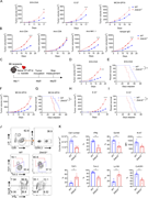
Figure 4. Loss of Zbtb32 inhibits the anti-tumor response of CD8 + T cells. (A) Tumor growth in WT and Zbtb32−/− mice after transplantation of 1 × 10⁶ B16, E.G7, or MC38 cells (n = 6 for each group). (B) Tumor growth in WT and Zbtb32−/− mice More about this image found in Loss of Zbtb32 inhibits the anti-tumor response of CD8 + T cel...
Images
Zbtb32 overexpression enhances CD8+T cell anti-tumor rejection. (A) Quantifications of specific genes expressions in WT, Zbtb32 OE activated CD8+ T cells measured by RT-qPCR (n = 3 for each group). (B) Schematic diagram of the transfer with 7 × 10⁵ transduced OT-I or P14 cells on day 8. (C) Tumor growth in mice transferred with 7 × 10⁵ activated Zbtb32 OE or RV control CD8+ OT-I cells on 8 days after 1 × 10⁶ B16-OVA cells inoculation (n = 6 for each group). (D) Survival rate in mice transferred with 7 × 10⁵ activated Zbtb32 OE or RV control CD8+ OT-I cells on day 8 after 1 × 10⁶ B16-OVA cells inoculation (n = 6 for each group). (E) Representative plots of TCF1 and Tim-3, IFNγ and GzmB in RV control, and Zbtb32 OE CD8+ OT-I TILs in B16-OVA TME. (F) Quantifications of specific molecules in RV control and Zbtb32 OE CD8+ OT-I TILs in B16-OVA TME (n = 6 for each group). Data are shown as means ± SEM; ns, not significant; *P < 0.05, **P < 0.01, ***P < 0.001, and ****P < 0.0001 by unpaired (A and F) two-tailed Student’s t test, Bonferroni-corrected two-way ANOVA (C), and Mantel–Cox test (D). Data shown in all graphs are a representative of three independent experiments.
Published: 06 February 2026
Figure 5. Zbtb32 overexpression enhances CD8 + T cell anti-tumor rejection. (A) Quantifications of specific genes expressions in WT, Zbtb32 OE activated CD8+ T cells measured by RT-qPCR (n = 3 for each group). (B) Schematic diagram of the More about this image found in Zbtb32 overexpression enhances CD8 + T cell anti-tumor rejecti...
Images
The impaired tumor control as a result of Zbtb32 deficiency in T cells is rescued in ICB treatment. (A) Schematic diagram of anti–PD-1 treatment assay in B16-OVA tumor-bearing mice, transferred with 3 × 10⁵ WT or 3 × 10⁵Zbtb32−/− naïve OT-I cells before 1 × 10⁶ B16-OVA cells inoculation. (B) Tumor growth in mice transplanted with 1 × 10⁶ B16-OVA cells after the transfer of 0.3 × 10⁶ naïve WT or Zbtb32−/− OT-I cells, with or without anti–PD-1 treatment (n = 6 for each group). (C) Comparison of tumor volumes in WT and Zbtb32−/− mice treated with ICB with those in untreated mice. (D) Representative plots of TCF1 and Tim-3, Ly108, and Cx3CR1 expression in WT and Zbtb32−/− OT-I TILs with or without anti-PD1 treatment. (E) Expressions of TCF1 and Tim-3, Ly108, and Cx3CR1 in WT and Zbtb32−/− OT-I TILs with or without anti–PD-1 treatment (n = 6 for each group). (F) Quantifications of cell number, Tpex and Ttex cell number in WT and Zbtb32−/− OT-I in TME or DLN with or without anti-PD1 treatment (n = 6 for each group). (G) MFI for Ki-67 in WT and Zbtb32−/− OT-I in TME with or without anti-PD1 treatment. (H) Representative plots of IFNγ and GzmB expression in WT and Zbtb32−/− OT-I TILs with or without anti-PD1 treatment. (I) Expressions of IFNγ, GzmB, MFI of Ki-67 and IL-2 in WT and Zbtb32−/− OT-I in TME with or without anti-PD1 treatment (n = 6 for each group). (J) Expression of TCF1 and IFNγ in WT and Zbtb32−/− OT-I in DLN with or without anti–PD-1 treatment (n = 6 for each group). Data are shown as means ± SEM; ns, not significant; *P < 0.05, **P < 0.01, ***P < 0.001 and ****P < 0.0001 by Bonferroni-corrected two-way ANOVA (B), unpaired two-tailed Student’s t test (C, E, F, I, and J). Data shown in all graphs are a representative of two independent experiments.
Published: 06 February 2026
Figure 6. The impaired tumor control as a result of Zbtb32 deficiency in T cells is rescued in ICB treatment. (A) Schematic diagram of anti–PD-1 treatment assay in B16-OVA tumor-bearing mice, transferred with 3 × 10⁵ WT or 3 × 10⁵Zbtb32−/− naïve More about this image found in The impaired tumor control as a result of Zbtb32 deficienc...
Images
Zbtb32 transcriptionally regulates tumor-infiltrating CD8+T cell function. (A) Schematic diagram of the co-transfer of 1.5 × 10⁵ WT and 1.5 × 10⁵Zbtb32−/− naïve OT-I cells prior to 1 × 10⁶ B16-OVA inoculation (n = 6 for each group). (B) Representative FACS plots of TCF1 and Tim-3, IFNγ, and GzmB expression in WT and Zbtb32−/− OT-I TILs. (C) Quantifications of the cell ratio and specific molecules of WT and Zbtb32−/− OT-I TILs. (D) Heatmaps showing relative expressions of different signature genes in WT and Zbtb32−/− OT-I TILs. (E) Volcanic plot showing expression levels of DEGs between WT and Zbtb32−/− OT-I TILs. (F) Lists of top 20 KEGG biological pathways for DEGs that were downregulated in Zbtb32−/− OT-I TILs. (G) GSEA analysis for comparing the enrichment DEGs in WT and Zbtb32−/− OT-I TILs (GEO accession: GSE114631). (H) GSEA analysis for a comparison of the enrichment DEGs in Tpex and Ttex CD8+ T cell subsets in WT and Zbtb32−/− OT-I TILs (GEO accession: GSE114631). Data are shown as means ± SEM; ns, not significant; **P < 0.01 and ****P < 0.0001 by paired two-tailed Student’s t test (C). Data shown are a representative of at least three independent experiments. NES, normalized enrichment score.
Published: 06 February 2026
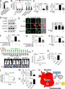
Figure 7. Zbtb32 transcriptionally regulates tumor-infiltrating CD8 + T cell function. (A) Schematic diagram of the co-transfer of 1.5 × 10⁵ WT and 1.5 × 10⁵Zbtb32−/− naïve OT-I cells prior to 1 × 10⁶ B16-OVA inoculation (n = 6 for each More about this image found in Zbtb32 transcriptionally regulates tumor-infiltrating CD8 + T ...
Images
Zbtb32 regulates the epigenetic landscape of CD8+TILs. (A) Distribution of differential chromatin accessible regions in WT and Zbtb32−/− OT-I TILs across the genome. (B) Enrichment of known TF-binding motifs in chromatin accessible regions between WT and Zbtb32−/− OT-I TILs. (C) Volcanic plot showing differential chromatin accessible regions between WT and Zbtb32−/− OT-I TILs. (D) Lists of top 20 GO enrichment pathways for differential chromatin accessible genes in WT and Zbtb32−/− OT-I TILs. (E) Chromatin accessible peaks at several classic gene loci in WT and Zbtb32−/− OT-I TILs aligned with assay for ATAC-seq tracks of Ttex and Tpex TILs from B16-OVA tumor. Binding peaks on Prdm1, Batf, Id2, Havcr2, Il2rb, Gzmb, and Gzmk loci are shown (GEO accession: GSE123236). (F) PSEA results for a comparison of the enrichment of epigenetic signature peaks in Tpex and Ttex CD8+ T cell subsets in WT and Zbtb32−/− OT-I TILs (GEO accession: GSE123236). PSEA, peak set enrichment analysis.
Published: 06 February 2026
Figure 8. Zbtb32 regulates the epigenetic landscape of CD8 + TILs. (A) Distribution of differential chromatin accessible regions in WT and Zbtb32−/− OT-I TILs across the genome. (B) Enrichment of known TF-binding motifs in chromatin More about this image found in Zbtb32 regulates the epigenetic landscape of CD8 + TILs. (A) ...
Images
Zbtb32 and Bcl6 exert antagonistic functions in tumor-specific CD8+T cells. (A) Consensus binding sequences of Zbtb32 and Bcl6. (B) Quantification of the ratios of ChIP-seq overlapped peaks bound by Zbtb32 and specific TFs in total Zbtb32-binding peaks in CD8+ T cells. (C) Heatmaps of read density profiles of Zbtb32 and other TFs ChIP-seq in CD8+ T cells surrounding Zbtb32-binding peaks, respectively. (D) Zbtb32, with or without Bcl6 overexpression, and Bcl6 occupancy at several classic gene loci were aligned with assay for ATAC-seq tracks of WT and Zbtb32−/− from B16-OVA tumor (GEO accession: GSE123236). The binding peaks on Prdm1, Batf, Id2, Havcr2, Il2rb, Gzmb, and Gzmk loci are shown. (E) ChIP-qPCR results showing the deposition of Zbtb32 at specific gene loci in CD8+ T cells following 4-day activation in vitro with or without Bcl6 overexpression. The detected sites are labeled by blue arrow in Fig. S5 H (n = 3 for each group). (F) Schematic diagram of the transfer with 7 × 10⁵ transduced OT-I cells on day 8. (G) The tumor growth in mice transferred with 0.7 × 10⁶ activated CD8+ T cells with Zbtb32, Zbtb32-B, Bcl6, or Bcl6-Z overexpression on day 8 after inoculation with 1 × 10⁶ B16-OVA cells (n = 6 for each group). (H) Schematic diagram of the co-transfer with 1.5×10⁵ WT and 1.5 × 10⁵Zbtb32−/− naïve OT-I cells. (I) Quantifications of relative cell ratios in Zbtb32, Zbtb32-B, Bcl6, and Bcl6-Z OE activated CD8+ T cells in comparison with RV control CD8+ T cells, co-transferred into recipients on day 8 after 1 × 10⁶ B16-OVA cells inoculation (n = 4 for each group). (J) Quantifications of specific molecules of transduced and RV control OT-I TILs in a co-transfer assay (n = 4 for each group). (K) ChIP-qPCR results demonstrating the deposition of NCoR1 at Prdm1 gene locus in CD8+ T cells following 4-day activation in vitro with or without Bcl6 overexpression (n = 3 for each group). GEO accession: GSE182034. Data are shown as means ± SEM; ns, not significant; *P < 0.05, **P < 0.01, and ***P < 0.001 by paired (J) and unpaired (E, I, and K) two-tailed Student’s t test and Bonferroni-corrected two-way ANOVA (G). Data shown are a representative (E, G, I, and J) or a pool (K) of two independent experiments. ATAC, transposase-accessible chromatin; Zbtb32-B, Zbtb32 with the Bcl6 BTB domain; Bcl6-Z, Bcl6 with the Zbtb32 BTB domain.
Published: 06 February 2026
Figure 9. Zbtb32 and Bcl6 exert antagonistic functions in tumor-specific CD8 + T cells. (A) Consensus binding sequences of Zbtb32 and Bcl6. (B) Quantification of the ratios of ChIP-seq overlapped peaks bound by Zbtb32 and specific TFs in More about this image found in Zbtb32 and Bcl6 exert antagonistic functions in tumor-specific CD8 + ...
Images
Zbtb32 upregulates Id2 to enhance CD8+T cell–mediated tumor rejection. (A) Fold changes in the expression levels of different TFs in DEGs in Zbtb32−/− and WT CD8+ TILs. (B) The occupancies of Zbtb32 and Bcl6 at the Id2 gene locus using ChIP-seq datasets. (C) ChIP-qPCR data showing the deposition of Zbtb32 at Id2 locus in CD8+ T cells in vitro activated for 4 days with or without Bcl6 overexpression. The detected sites are labeled by blue arrows in Fig. 7 B (n = 3 for each group). (D) Quantifications of Id2 expression in Bcl6-, Zbtb32-, double OE, and RV control activated CD8+ T cells measured by RT-qPCR (n = 3 for each group). (E)Id2 expression in WT and Zbtb32 OE CD8+ T cells (left panel) and Zbtb32 expression in WT and Id2 OE CD8+ T cells (right panel) measured by RT-qPCR. (F) Schematic diagram of Id2 rescue assay in B6 mice, transferred with 7 × 10⁵ transduced CD8+ T cells on day 8 after inoculation of 1 × 10⁶ B16-OVA cells. (G) Tumor growth in WT and Zbtb32−/− mice transferred with activated WT or Zbtb32−/− CD8+ OT-I cells with or without Id2 overexpression (n = 6 for each group). (H) Schematic diagram of Id2 rescue assay in B6 mice, co-transferred with 5 × 10⁵ transduced CD8+ T cells on day 8 after inoculation of 1 × 10⁶ B16-OVA cells. (I) Representative plots and quantifications of TCF1 and Tim-3 expressions in co-transferred WT and Zbtb32−/− CD8+ TILs with or without Id2 overexpression (n = 4 for each group). (J) MFI and quantification of GzmB, IFNγ, and Ki-67 in WT and Zbtb32−/− CD8+ OT-I TILs with or without Id2 overexpression (n = 4 for each group). Data are shown as means ± SEM; ns, not significant; *P < 0.05, **P < 0.01, ***P < 0.001, and ****P < 0.0001 by paired (I) and unpaired (C, D, E, and J) two-tailed Student’s t test and Bonferroni-corrected two-way ANOVA (G). Data shown are a representative (C–J) of two independent experiments.
Published: 06 February 2026
Figure 10. Zbtb32 upregulates Id2 to enhance CD8 + T cell–mediated tumor rejection. (A) Fold changes in the expression levels of different TFs in DEGs in Zbtb32−/− and WT CD8+ TILs. (B) The occupancies of Zbtb32 and Bcl6 at the Id2 gene More about this image found in Zbtb32 upregulates Id2 to enhance CD8 + T cell–mediated tumor ...
Journal Articles
Images
Images
PTL suppresses brain tumor progression by blocking nicotine-induced M2 microglia polarization. (A) Human microglia cells (HMC3) with the Arg1 reporter plasmid were cultured in the presence or absence of compounds that were identified as the top three most effective inhibitors for Arg1 during our initial screening (see Materials and methods). After 48 h of incubation, luciferase reporter activity was measured (n = 4/group). (B) Expression of surface markers of M1/M2 microglia was examined by qRT-PCR after microglial cells were treated with or without nicotine plus PTL (n = 4/group). (C) The same set of samples in B was evaluated for quantification of Iba1+/CD11b+ (M1) cells and Iba1+/CD206+ (M2) cells by FACS (n = 4/group). (D) The same set of samples in C was examined for quantification of the protein expression of JAK2 and STAT3 by Western blot. (E) Human microglial (HMC3) cells (green) with or without nicotine treatment (1 µM) in the presence or absence of PTL (1 µM) were incubated with PKH26-labeled H2030BrM cells (red) for 24 h and photographed (left panels), followed by measurement of the microglial phagocytic activity (right panel; n = 4/group). Scale bar, 10 µm. (F) CM was prepared from human microglia (HMC3) treated with or without nicotine and PTL. The CM was added to the culture of H2030BrM, and cells were incubated for 48 h followed by evaluation of CSC population by FACS. Non-nicotine, nicotine, or Nico+PTL CM: microglia were treated with PBS, nicotine, or nicotine plus PTL for 24 h. They were then washed twice with PBS and incubated in the fresh DMEM/F12 medium supplemented with 2% FBS for a further 24 h (n = 4/group). (G) For the same set of samples as F, colony-forming ability was also measured (n = 4/group). (H) Human microglia were treated with or without nicotine (1 µM) in the presence or absence of PTL for 24 h, followed by assessment of the expression of CCL20 by qRT-PCR (n = 4/group). (I) The mouse lung cancer cells, LL/2, were intracardially injected into wild-type BALB/c mice. After 3 d of intracranial transplantation of LL/2 cells, mice received nicotine (1 mg/kg) plus PTL (1 mg/kg) by an intraperitoneal injection every 3 d for 40 d. Upper panel: BLI images of representative mice from each experimental group at day 40. Lower panel: total photon flux of ex vivo brain metastatic lesions was measured by BLI at the endpoint (day 40; n = 9/group). (J) Quantitative data of BLI in the brain regions are shown (n = 9/group). (K) Ex vivo signals in the whole brains at the end point were quantified. (L) The Kaplan–Meier analysis of brain metastasis–free survival was performed (n = 9/group). (M and N) Metastatic brain tumors in I were isolated from the brain and were examined by FACS for M2 (M) and M1 (N) microglial polarization (n = 9/group). (O) A proposed model illustrating a nicotine-induced brain metastasis (n = 9/group). The data are presented as the mean ± SD. *, P < 0.05; **, P < 0.01; and ***, P < 0.001.
Published: 05 February 2026
Figure 6. PTL suppresses brain tumor progression by blocking nicotine-induced M2 microglia polarization. (A) Human microglia cells (HMC3) with the Arg1 reporter plasmid were cultured in the presence or absence of compounds that were identified More about this image found in PTL suppresses brain tumor progression by blocking nicotine-induced M2 micr...
Images
Journal Articles
Journal Articles
Journal Articles
Images
Characterizing pre-plasma B cells through scRNA-seq analysis. (A) UMAP visualization of 10 GC B cell clusters. (B) UMAP visualization of Irf4 expression and enrichment of PC signature in clusters as in A (left). Dot plot showing the expression of Irf4 and enrichment of PC signature in clusters as in A (right). (C) Pseudotime analysis of DZ, LZ, LZ7 GC B cells, and PCs. (D) UMAP visualization of the expression of CD205 (left) and CD83 (right) in clusters as in A. (E) Dot plot of the expression of CD205 in clusters as in A. (F) Flow cytometry profile showing CD205+ CD83+ GC B cells. (G) Flow cytometry profile showing the relationship between CD138+ GC B cells and CD205hi GC B cells. (H) Flow cytometry analysis of forward scatter in CD205low, LZ, and CD205hi GC B cells. (I) Representative transmission electron microscopy of CD205low, CD205hi GC B cells, and PCs (left) and the ratio of ER area to cytosol area in indicated subsets (right). Scale bars, 1 and 0.5 μm in original and magnified images, respectively. (J) Output of in vitro GC B cell culture system on CD205low, LZ, and CD205hi GC B cells from LCMV-c13–infected mice. (K) Schematic of the experiment (left). Gating strategies shown by flow cytometry profile (middle). The total number of splenic PCs in mice (right). Data represent two (I and J) or three (H and K) independent experiments. Five to ten mice were used for each experiment (H, I, and J). Five to ten mice were used for the donors, and three to four mice were used for the recipients (K). Data are the mean ± SD; NS, not significant, **P < 0.01, ***P < 0.001. One-way ANOVA (H, I, and J) was used for data analysis. UMAP, Uniform Manifold Approximation and Projection.
Published: 28 January 2026
Figure 1. Characterizing pre-plasma B cells through scRNA-seq analysis. (A) UMAP visualization of 10 GC B cell clusters. (B) UMAP visualization of Irf4 expression and enrichment of PC signature in clusters as in A (left). Dot plot showing the More about this image found in Characterizing pre-plasma B cells through scRNA-seq analysis. (A) UMAP vis...
Images
Kdm6b is upregulated in prePCs. (A) Genome track view of the Irf4 locus showing H3K27me3 (blue, above the line) and H3K4me3 (red, below the line) peaks. (B) Expression of Ezh2, Kdm6a, and Kdm6b in indicated clusters visualized using UMAP. Dot plot showing the expression of Ezh2, Kdm6a, and Kdm6b in clusters. (C) Diagram of Efnb1CreERT2/+; Rosa26LSL-YFP/+ mice and the schematic of the experiment (left), flow cytometry profile showing gating for YFP+ cells in GC B cells, MBCs, and PCs (middle), and the frequency of YFP+ cells in these cells (right). (D) Diagram of Kdm6bDre+; Efnb1CreERT2/+; Rosa26CAG-LSL-RSR-tdTomato mice and the schematic of the experiment (left), flow cytometry profile showing gating for tdTomato+ cells in GC B cells, MBCs, and PCs (middle), and the frequency of tdTomato+ cells in these cells (right). (E)Kdm6a and Kdm6b mRNA levels in follicular B (FoB) cells or GC B cells after 6-h stimulation with anti-IgM, anti-CD40, both anti-IgM and anti-CD40, or respective controls. (F)Nr4a1, Kdm6a, and Kdm6b mRNA levels in Nur77-GFP− and Nur77-GFP+ GC B cells in Nur77 reporter mice. Data represent two independent experiments (E and F). Six mice were used in C, and seven mice were used in D. Data are the mean ± SD; NS, not significant, **P < 0.01,***P < 0.001. Two-tailed unpaired Student’s t test (E and F) was used for data analysis. MBCs, memory B cells.
Published: 28 January 2026
Figure 2. Kdm6b is upregulated in prePCs. (A) Genome track view of the Irf4 locus showing H3K27me3 (blue, above the line) and H3K4me3 (red, below the line) peaks. (B) Expression of Ezh2, Kdm6a, and Kdm6b in indicated clusters visualized using More about this image found in Kdm6b is upregulated in prePCs. (A) Genome track view of the Irf4...