
Skip Nav Destination
Close Modal


1-20 of 61
Follow your search
Access your saved searches in your account
Would you like to receive an alert when new items match your search?
1
Journal Articles
Journal:
Journal of Experimental Medicine
J Exp Med (2017) 215 (1): 35–49.
Published: 14 December 2017
in Organ-specific lymphatic vasculature: From development to pathophysiology
> Journal of Experimental Medicine
Published: 14 December 2017
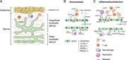
Figure 1. Organization and function of dermal lymphatic vasculature. (A) Skin LVs are organized in superficial and deep lymphatic plexuses. Superficial LVs are mostly capillaries, whereas deep lymphatic plexus contain collecting LVs draining to More about this image found in Organization and function of dermal lymphatic vasculature. (A) Skin LVs ar...
in Organ-specific lymphatic vasculature: From development to pathophysiology
> Journal of Experimental Medicine
Published: 14 December 2017
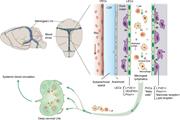
Figure 2. Meningeal lymphatic vasculature and brain mural LECs. Localization and distribution of LVs in dura matter of mouse brain. Cerebrospinal fluid (CSF) and interstitial fluid of brain parenchyma drain into meningeal LVs and reach the deep More about this image found in Meningeal lymphatic vasculature and brain mural LECs. Localization and dis...
in Organ-specific lymphatic vasculature: From development to pathophysiology
> Journal of Experimental Medicine
Published: 14 December 2017
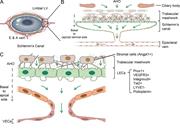
Figure 3. Schlemm's canal. (A) SC is an endothelium-lined channel that encircles the cornea and provides an exit route for aqueous humor. (B) Aqueous humor is produced from the ciliary body and drained into aqueous and episcleral veins through More about this image found in Schlemm's canal. (A) SC is an endothelium-lined channel that encircles the...
in Organ-specific lymphatic vasculature: From development to pathophysiology
> Journal of Experimental Medicine
Published: 14 December 2017
Figure 4. LN lymphatic vasculature. (A) Afferent LVs deliver lymph carrying antigens and immune cells to the LN SCS. From the SCS, lymph flows to the cortical and medullary sinuses and exits via efferent LVs. SCS “ceiling” LECs (cLECs) express More about this image found in LN lymphatic vasculature. (A) Afferent LVs deliver lymph carrying antigens...
in Organ-specific lymphatic vasculature: From development to pathophysiology
> Journal of Experimental Medicine
Published: 14 December 2017
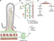
Figure 5. LVs of the small intestine. (A) Intestinal lacteals are positioned in the middle of intestinal villi. Smooth muscle cells in the villi are closely associated with lacteals, and their contractions promote lymph uptake and transport. More about this image found in LVs of the small intestine. (A) Intestinal lacteals are positioned in the ...
Journal Articles
In Special Collection:
Vascular Biology and Human Disease
Journal:
Journal of Experimental Medicine
J Exp Med (2017) 214 (12): 3645–3667.
Published: 15 November 2017
Includes: Supplementary data
Published: 15 November 2017
Figure 1. Postnatal development of the meningeal lymphatic network. (A and B) Schematic illustration showing meningeal LVs (green) superimposed on the corresponding anatomical structures at different postnatal (P) days (A) and the direction of More about this image found in Postnatal development of the meningeal lymphatic network. (A and B) Schema...
Published: 15 November 2017
Figure 2. LVs in spinal meninges. (A) Schematic illustration of meningeal LVs (green) attached to the ventral and dorsal sides of the cranium and spinal canal after removal of the brain and spinal cord. SN; spinal nerve. (B) Transverse section More about this image found in LVs in spinal meninges. (A) Schematic illustration of meningeal LVs (green...
Published: 15 November 2017
Figure 3. LV exit from the spinal canal along the spinal nerves and BVs. (A) Schematic transverse view of the spinal cord and its blood (red) and lymphatic (green) vessels. (B) Transverse section of spinal cord with a close-up showing the exit More about this image found in LV exit from the spinal canal along the spinal nerves and BVs. (A) Schemat...
Published: 15 November 2017
Figure 4. Sprout extension and fusion of cell clusters in meningeal lymphangiogenesis. (A) LYVE1 staining of LVs developing around the PPA. (B) LYVE1+/CD206+ macrophage-like cells around the SSS at P16. (C) CD206 immunostaining around the MMA. More about this image found in Sprout extension and fusion of cell clusters in meningeal lymphangiogenesis...
Published: 15 November 2017
Figure 5. VEGF-C, but not VEGF-D, is essential for normal meningeal LV development. (A and B) LYVE1 (gray) and PROX1 (green) staining of the FM area in P12 VegfcLacZ/+ (n = 3, 3; P = 0.0429; A) and Vegfd−/− mice and their littermate controls (n More about this image found in VEGF-C, but not VEGF-D, is essential for normal meningeal LV development. ...
Published: 15 November 2017
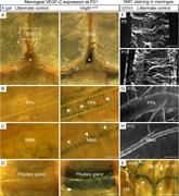
Figure 6. Smooth muscle cells provide a vascular source of VEGF-C for meningeal LVs. (A–E) β-Galactosidase staining of meningeal tissue showing VEGF-C expression around the TS and SSS and in the pineal gland (asterisks; A), PPA (B), MMA (C), More about this image found in Smooth muscle cells provide a vascular source of VEGF-C for meningeal LVs. ...
Published: 15 November 2017
Figure 7. VEGFR-3 is essential for meningeal LV development. (A and B) Comparison of dural LYVE1 staining in P21 mice deleted of Vegfr3 (Vegfr3iΔR26, n = 4) and their littermate controls (Vegfr3flox/flox, n = 9) around the TS (A) and MMA (B). More about this image found in VEGFR-3 is essential for meningeal LV development. (A and B) Comparison of...
Published: 15 November 2017
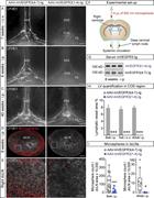
Figure 8. Meningeal LV growth in response to AAV–mVEGF-C. (A–E) Analysis of meningeal LVs in mice injected i.c.v. with AAV–mVEGF-C (n = 9) or AAV without payload (empty-AAV; n = 9). LYVE1 staining of the CN II (A) and COS area (B), and More about this image found in Meningeal LV growth in response to AAV–mVEGF-C. (A–E) Analysis of meningea...
Published: 15 November 2017
Figure 9. VEGFR-3 signaling is required for LV maintenance in adult meninges. (A and B) Comparison of LYVE1 staining around the TS (A) and PPA (B) in Rosa26-Vegfr3flox/flox (n = 4) and Vegfr3iΔR26 mice (n = 4) 20 wk after tamoxifen More about this image found in VEGFR-3 signaling is required for LV maintenance in adult meninges. (A and...
Published: 15 November 2017
Figure 10. Regression of meningeal LVs decreases drainage of i.c.v.-injected microspheres. (A–C) Representative images of LYVE1-stained LVs around the COS 7 wk after i.c.v. injection (A; n = 6, 6), 8 wk after i.p. injection (B; n = 6, 6), and 40 More about this image found in Regression of meningeal LVs decreases drainage of i.c.v.-injected microsphe...
Journal Articles
Journal:
Journal of Experimental Medicine
J Exp Med (2017) 214 (11): 3151–3169.
Published: 23 October 2017
in Alzheimer’s disease: A matter of blood–brain barrier dysfunction?
> Journal of Experimental Medicine
Published: 23 October 2017
Figure 1. Contribution of BBB breakdown and dysregulated BBB transport to AD pathophysiology based on findings in animal models, as shown in Tables 1 and 2 . BBB breakdown (left) leads to perivascular accumulation of blood-derived More about this image found in Contribution of BBB breakdown and dysregulated BBB transport to AD pathophy...
Journal Articles
In Special Collection:
Vascular Biology and Human Disease
Journal:
Journal of Experimental Medicine
J Exp Med (2017) 214 (11): 3331–3346.
Published: 28 September 2017
Includes: Supplementary data
1

