NBAS deficiency is a multisystem disorder, but its immunologic manifestations and underlying mechanisms remain poorly defined. Here, we expand the genotypic and phenotypic spectrum of NBAS deficiency through four patients carrying five loss-of-function NBAS mutations. Minigene assays and RNA analysis showed that these splicing or missense mutations resulted in aberrant alternative splicing. Clinically, the patients showed early-onset recurrent or persistent cytopenias and immune dysregulation, together with severe infections, inflammatory activation, and multi-organ involvement, including renal and neurologic manifestations. Transcriptomic analysis revealed overactivation of type I interferon signaling pathways compared with healthy controls, and single-cell RNA sequencing demonstrated that this upregulated inflammation was predominantly driven by myeloid cells. Together, these findings identify NBAS deficiency as a cause of immune dysregulation characterized by myeloid-driven type I interferonopathy, expand the clinical spectrum of NBAS-related disorders, and highlight the critical role of NBAS in maintaining immune homeostasis.
Introduction
Immune dysregulation diseases comprise a heterogeneous group of disorders characterized by aberrant immune responses and disrupted homeostasis. These conditions arise from defects in immune regulatory pathways, in which the genetic causative components play an important role in pathogenic mechanisms (1, 2).
Neuroblastoma amplified sequence (NBAS) encodes a subunit of the NRZ tethering complex, which is involved in Golgi-to-ER retrograde transport with the COPI complex. NBAS is a component of the trimeric NRZ complex together with RINT1 and ZW10, which interacts with ER SNAREs syntaxin 18, BNIP1, p31, and Sec22b and regulates the coupling and fusion of COPI with the ER. NBAS serves as a link between p31 and ZW10-RINT1 with its N-terminal and C-terminal region, respectively. NBAS depletion impairs COPI-dependent Golgi-to-ER retrograde transport of proteins (3, 4). Defects in retrograde transport from the Golgi back to the ER, which is essential for retrieving cycling and escaped resident ER proteins, have been described to cause human diseases. Coatomer subunit α (COPA) is a subunit of COPI complex, and its mutation causes autoimmune disease with elevated type I interferon (IFN-I) signaling (5, 6, 7, 8, 9). NBAS has also been reported to regulate nonsense-mediated decay (NMD) in human cells. Depletion of NBAS results in a significant increase in mRNA levels of NMD genes (10).
NBAS mutations have been reported to cause two major autosomal recessive disorders covering multiple systems, including short stature, optic nerve atrophy, and Pelger–Huët anomaly (SOPH) (OMIM #614800) (11) characterized by severe postnatal growth retardation, facial dysmorphism, short stature, and Pelger–Huët anomaly of leucocytes; and infantile liver failure syndrome 2 (ILFS2) (OMIM #616483) characterized by recurrent fever with acute liver failure (12). The development of hemophagocytic lymphohistiocytosis (HLH) has also been described as a clinical manifestation associated with NBAS mutations (13). Notably, ∼44% of patients with NBAS mutations exhibit immunologic impairment. However, previous investigations have been largely limited to characterizing quantitative and functional defects in specific lymphocyte subsets, such as natural killer (NK) and B cells (14, 15). The broader transcriptional landscapes and specific inflammatory signaling pathways across distinct cell lineages, particularly the contribution of myeloid cells to systemic inflammation, remain largely unexplored.
In this study, we report four patients with genetically confirmed NBAS deficiency who exhibited early-onset recurrent or persistent cytopenias, immune dysregulation, infection susceptibility, inflammatory activation, and multisystem involvement, including renal and neurologic manifestations. These findings expand the phenotypic spectrum of NBAS-related disease beyond its classical presentations. Using bulk RNA sequencing (RNA-seq) and single-cell RNA sequencing (scRNA-seq), we demonstrate for the first time that NBAS deficiency drives an elevation of IFN-I signaling, providing novel insights into the critical role of NBAS in maintaining immune homeostasis.
Results
Clinical information of patients
Four Chinese patients diagnosed with NBAS deficiency were included in this study (Table S1). Patient 1 (P1) was a male patient who initially presented with thrombocytopenia at 3 years of age, together with autoimmune features including positive antinuclear antibody (ANA) and anti-SSA antibody. His disease subsequently evolved into recurrent cytopenia and multisystem involvement. At ∼7 years of age, he developed his first ischemic stroke, followed by recurrent cerebral infarctions and progressive renal involvement with persistent proteinuria. Renal biopsy showed secondary membranous nephropathy-like changes with IgM deposition, and a later biopsy demonstrated membranoproliferative-like glomerular lesions considered related to immune complex and thrombotic microangiopathy-like injury. During the later disease course, he developed chronic kidney disease, severe recurrent infections, necrotizing oral/facial soft-tissue lesions with loss of palatal and gingival tissue, persistent anemia and thrombocytopenia, and marked lymphopenia. He received multiple immunomodulatory and supportive interventions, but these treatments resulted in only partial and transient improvement, and the disease remained relapsing and progressive. He later died from massive gastrointestinal bleeding.
Patient 2 (P2) was a male patient with early-onset renal and hematologic disease. He developed nephrotic syndrome–like manifestations in early childhood, with recurrent lower limb and eyelid edema and abnormal urinalysis. From ∼6 to 7 years of age, he had recurrent cytopenias involving platelets, hemoglobin, and leukocytes. The cytopenias were partially responsive to corticosteroids but repeatedly relapsed during tapering. Later hematologic evaluation documented severe thrombocytopenia and neutropenia. He subsequently developed nephrotic-range proteinuria, hypoalbuminemia, autoimmune hypothyroidism, and hypogammaglobulinemia. During a later admission, he developed severe pneumonia with respiratory failure, with respiratory syncytial virus positivity and molecular evidence of Pneumocystis jirovecii and cytomegalovirus infection. He received corticosteroids and other immunomodulatory therapies, together with supportive care. Edema and respiratory manifestations improved after treatment, and proteinuria decreased; however, the hematologic and renal disease followed a recurrent course of proteinuria.
Patient 3 (P3) was a 4-year-old girl who presented with thrombocytopenia for more than 1 mo, malnutrition, mild pallor, and hepatosplenomegaly. Initial laboratory testing showed anemia, thrombocytopenia, elevated inflammatory markers, liver dysfunction, hypokalemia, metabolic acidosis, and positive antinuclear antibody. During hospitalization, she fulfilled five HLH-2004 diagnostic criteria (16) and was clinically diagnosed with HLH, including recurrent fever >38.5°C, hepatosplenomegaly/splenomegaly, cytopenias affecting at least two lineages, bone marrow hemophagocytosis, and markedly elevated soluble IL-2 receptor/sCD25. Her cytopenias subsequently worsened, with severe thrombocytopenia and anemia, increased inflammatory cytokines, positive platelet antibody screening, and viral coinfection with cytomegalovirus and rhinovirus. She later deteriorated with difficult-to-control epistaxis, marked cytokine activation, and high proportions of activated CD8 T cells. After ruxolitinib-based treatment combined with corticosteroids and supportive care, bleeding and inflammatory activity improved, platelet counts recovered, C-reactive protein (CRP) normalized, and soluble IL-2 receptor decreased. After discharge, she underwent hematopoietic stem cell transplantation at another hospital and died from transplant-related complications.
Patient 4 (P4), the younger sister of P3, was a 2-year-old girl who presented with pallor for more than 6 mo. Unlike P3, she had a milder hematologic phenotype characterized mainly by anemia, leukopenia, and neutropenia, while platelet counts were preserved and no frank HLH crisis was documented. Bone marrow evaluation showed abnormal hematopoiesis with decreased granulopoiesis, active erythropoiesis, and no evidence of leukemia. She also had mild inflammatory and immune activation markers, including elevated soluble IL-2 receptor and CXCL9. She received anti-infective treatment, red blood cell transfusion, intravenous immunoglobulin, prednisone, and supportive care. Her anemia and general clinical condition improved, although neutropenia persisted.
Overall, all four patients showed early-onset and persistent or recurrent cytopenia as a prominent shared manifestation of NBAS deficiency. Patients in this study also exhibited immune dysregulation, including infection susceptibility, autoimmune features with positive autoantibodies and inflammatory activation, and multisystem involvement affecting the renal and neurologic systems. These clinical features, including antinuclear antibody (ANA)/anti-SSA positivity, immune complex–mediated glomerular disease, and recurrent childhood ischemic stroke, extended beyond the classical phenotypic spectrum of NBAS deficiency (Table 1).
Patients with the NBAS variants
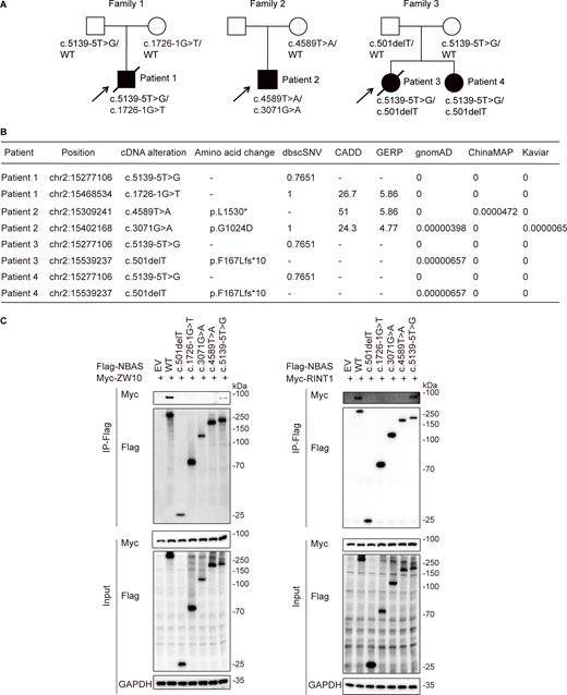
Whole-exome sequencing (WES) revealed the presence of five splicing or stop NBAS mutations in three families. P1 was compound-heterozygous for the c.5139-5T>G and c.1726-1G>T, and P2 was compound-heterozygous for the c.4589T>A (p.L1530*) and c.3071G>A (p.G1024D) NBAS (RefSeq: NM_015909.4) variants (Fig. 1 A and Fig. S1 A). P3 and P4 were compound-heterozygous for the c.5139-5T>G and c.501delT (p.F167Lfs*10) (Fig. 1 A). Among these mutations, only c.5139-5T>G and c.501delT have been previously reported (17, 18), while other mutations were novel. These mutations were rare and not present as homozygotes in public databases, including gnomAD, ChinaMAP, and Kaviar. In addition, these mutations were predicted to be deleterious by multiple software. The stop mutation c.4589T>A (p.L1530*) was a predicted loss-of-function variant with combined annotation-dependent depletion (CADD) Phred score of 51, and the splicing or missense mutations were predicted to be pathogenic with the CADD Phred score of 26.7 and 24.3, respectively (Fig. 1 B). Sanger sequencing validated the identified mutations and confirmed their compound-heterozygous state (Fig. S1 B). These data suggested that the clinical phenotypes in the three families were inherited recessively and segregated with the inheritance of NBAS, implying the potential pathogenic role of these NBAS variants.
NBAS splicing or missense mutations cause aberrant alternative splicing
As the mutations c.1726-1G>T and c.5139-5T>G were located in the intron of NBAS and the mutation c.3071G>A was located in the last nucleotide of the exon, these mutations were presumed to cause aberrant alternative splicing. In addition, these mutations were predicted to be splice altering variants with the dbscSNV value of 0.7651 or 1 (Fig. 1 B). To confirm the predicted impact of these mutations on NBAS gene splicing, we extracted and analyzed RNA from P1 and his parents. Sanger sequencing of the reverse transcription–PCR (RT-PCR) products showed that the mutation c.1726-1G>T caused the loss of the first two nucleotides of exon 17, introducing a premature stop codon (p.S576*), while the mutation c.5139-5T>G caused the deletion of exon 43 and the introduction of a premature stop codon (p.L1714Sfs*2) (Fig. S1, C–E). To verify the effect of mutation c.3071G>A, wild-type and mutant c.3071G>A genomic fragments of NBAS were cloned into the pCAS2 vector and HEK293T cells were transfected with the minigene. The mRNA sequence showed that the mutation c.3071G>A resulted in the deletion of exon 26 and introduction of a premature stop codon (p.L980*) (Fig. S1, F and G). As the C-terminal (amino acids 1,036–2,371) of NBAS is responsible for the interaction with ZW10-RINT1, and the truncated C-terminal NBAS has no interaction with ZW10 and RINT1 (3), we performed co-immunoprecipitation assays to directly evaluate whether these variants affect the interaction between NBAS and its binding partners. c.1726-1G>T, c.4589T>A, c.3071G>A, and c.501delT mutant NBAS proteins exhibited complete loss of interaction with ZW10 and RINT1, and c.5139-5T>G mutant NBAS proteins exhibited impaired interaction with ZW10 and RINT1, whereas the wild-type protein retained robust binding (Fig. 1 C). These results provide direct experimental evidence that the variants disrupt the formation of the NBAS–ZW10–RINT1 complex, supporting a loss-of-function mechanism.
Elevated inflammatory signals in patient’s peripheral blood mononuclear cells (PBMCs)
Considering the essential role of NBAS in COPI-dependent retrograde transport, it is likely that NBAS deficiency mimics the pathogenic mechanism of COPI dysfunction, potentially leading to the overactivation of the IFN-I pathway.
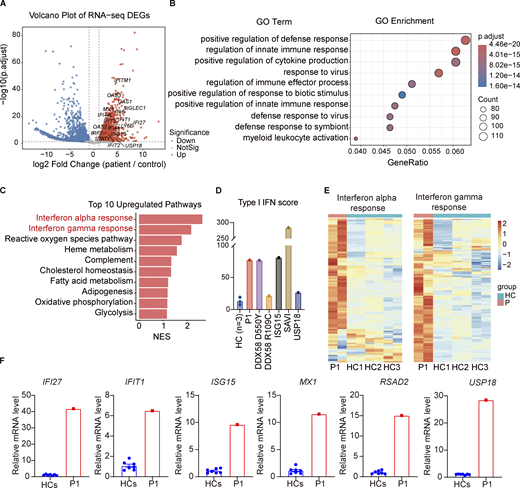
To verify whether NBAS deficiency is associated with the dysregulation of IFN-I production, we performed RNA-seq to profile the inflammatory transcriptome of P1 during persistent renal dysfunction and cytopenia. Immune-related genes including IFI44L, OAS1, OAS3, and SIGLEC1 were upregulated in the patient, and differentially expressed genes in the patient compared with healthy controls were enriched in the immune response–regulating signaling pathway and the regulation of innate immune pathway (Fig. 2, A and B). Gene set enrichment analysis (GSEA) enrichment showed the elevation of interferon alpha (IFN-α) and interferon gamma (IFN-ɣ) pathways in P1 (Fig. 2 C). Since the IFN-I score is considered as an indicator of autoimmune and autoinflammatory disorders, we compared the IFN-I score between healthy controls, P1, and reported cases of monogenic interferonopathies including DDX58 gain-of-function mutation (19, 20), ISG15 deficiency (21), STING gain-of-function mutation (22), and USP18 deficiency (23). The IFN-I score was markedly increased in P1 and fell within the range observed in patients with DDX58 mutations and ISG15 deficiency (Fig. 2 D). Heatmap analysis further confirmed that the transcriptional levels of several genes in IFN-α and IFN-ɣ pathways were elevated in P1 (Fig. 2 E). To validate these transcriptomic findings with laboratory tests, we performed quantitative real-time PCR (qPCR) on the PBMCs of P1. Consistent with the RNA-seq data, the qPCR results demonstrated a marked upregulation of interferon (IFN)-stimulated genes (ISGs), including IFI27, IFIT1, ISG15, MX1, RSAD2, and USP18 (Fig. 2 F). Together, these results indicated that inflammatory responses, especially IFN responses, were elevated in the NBAS-deficient patient.
Activated inflammation in patient’s bone marrow hematopoietic cells
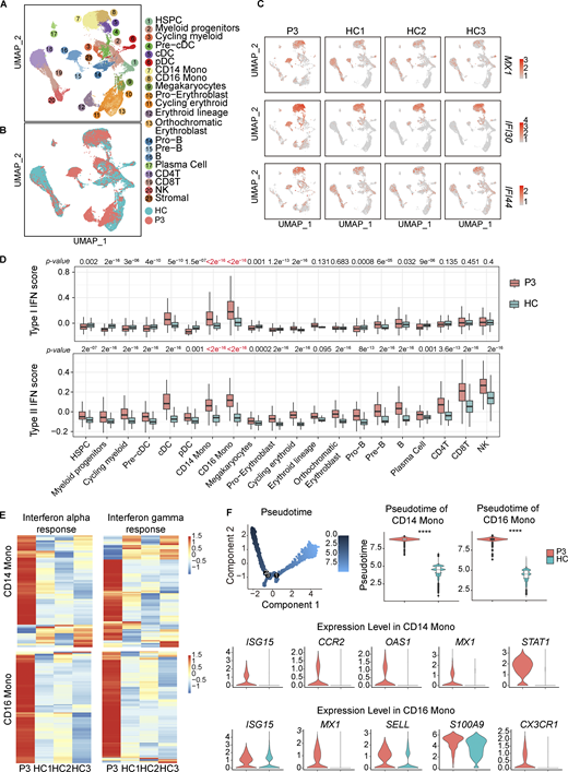
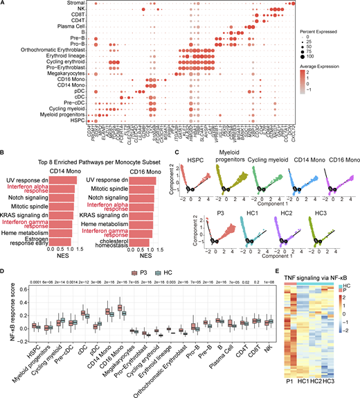
To elucidate the impact of NBAS deficiency on immune profile, we performed scRNA-seq on bone marrow hematopoietic cells isolated from P3 during transient partial hematologic improvement before a subsequent HLH inflammatory flare. Dimensionality reduction and clustering analysis identified 29 distinct clusters, which were annotated based on canonical marker gene expression (Fig. 3, A and B; and Fig. S2 A). Proinflammatory mediators and ISGs including MX1, IFI44, and IFI30 were transcriptionally upregulated in P3, especially in monocytes (Fig. 3 C). Analysis of IFN-I and type II IFN score confirmed the hyperinflammatory state in P3, particularly in myeloid cells (Fig. 3 D). GSEA and heatmaps revealed that gene sets, such as IFN-α pathway and IFN-ɣ pathway, were enriched in patient’s myeloid cells compared with healthy controls (Fig. S2 B and Fig. 3 E). Pseudotime trajectory analysis of hematopoietic stem and progenitor cells and myeloid subsets showed that CD14 and CD16 monocytes in the patient occupied a more advanced differentiation state and were more activated than healthy controls, confirmed by higher expression of inflammatory signature (Fig. 3 F and Fig. S2 C). Furthermore, NF-κB activation was also consistently observed in both P1 and P3 (Fig. S2, D and E). Collectively, these results demonstrated that NBAS deficiency drove a profound inflammatory response in bone marrow hematopoietic cells, which was predominantly sustained by the myeloid compartment.
Discussion
In this study, we report four patients carrying compound-heterozygous NBAS variants. These patients exhibited an underrecognized immune dysregulation phenotype characterized by early-onset persistent or recurrent cytopenia, systemic inflammatory activation, recurrent infections, autoimmune features, and renal and neurologic involvement. Through comprehensive transcriptomic analysis of PBMCs and scRNA-seq of bone marrow hematopoietic cells, we demonstrate for the first time that NBAS deficiency drives a profound elevation of IFN-I signaling. This finding is clinically corroborated by the therapeutic efficacy of ruxolitinib, a JAK inhibitor targeting this pathway, in P3. These findings not only expand the genotypic spectrum of NBAS variants but also define a novel immune dysregulation phenotype driven by myeloid hyperactivation.
NBAS deficiency has increasingly been recognized as an inborn error of immunity. Our patients shared previously recognized immunologic features of NBAS deficiency, including recurrent infections, hypogammaglobulinemia, and B and NK cell abnormalities. Beyond these established features, patients in our study exhibited an expanded phenotype characterized by autoimmunity with ANA and anti-SSA positivity and immune complex–mediated glomerular disease. These manifestations overlap with the clinical spectrum of type I interferonopathies (24) and may be related to the prominent monocyte IFN activation identified in our study. Thus, monocyte IFN activation might represent an additional inflammatory mechanism contributing to the expanded immune phenotype of NBAS deficiency.
Mechanistically, it is likely that this IFN signature in our patients may arise from a defect in retrograde transport, similar to that observed in COPA syndrome, in which defective COPI-mediated STING retrieval causes constitutive IFN-I activation (5, 7, 9, 25). As NBAS cooperates with COPI-dependent retrograde transport as part of the NRZ tethering complex, NBAS deficiency may similarly disturb trafficking homeostasis and promote IFN activation. DDX58-, ISG15-, and USP18-associated interferonopathies converge on IFN-I activation through different mechanisms, including enhanced RIG-I–mediated nucleic acid sensing (19, 20), impaired ISG15-dependent USP18 stabilization (21), or defective negative feedback downstream of IFNAR signaling (23). In contrast, NBAS deficiency may link impaired intracellular trafficking to monocyte-driven IFN activation, suggesting a trafficking-associated route to interferonopathy-like immune dysregulation.
Our findings have potential clinical implications, particularly for NBAS-deficient patients with prominent inflammatory activation. The identification of a prominent IFN-I signature suggests that JAK inhibitors, which block IFN signaling, may offer a targeted therapeutic approach for this specific subset of NBAS-deficient patients. The clinical improvement observed in P3 following treatment with ruxolitinib combined with corticosteroids supports the potential efficacy of JAK inhibitors.
Several limitations of this study should be acknowledged. First, because NBAS deficiency is rare, the study size was small; therefore, the expanded immune phenotype described here will require validation in additional patients. Second, transcriptomic analyses support IFN-I activation as a feature of NBAS deficiency, longitudinal samples across active disease and remission will be needed to determine the persistence and dynamics of this IFN signature. Finally, the therapeutic response to ruxolitinib is based on a single patient. Future clinical evaluation of JAK inhibition in more NBAS-deficient patients will be essential to establish the efficacy and safety of this treatment approach.
In conclusion, we identify five NBAS mutations in four patients with a previously underrecognized immune dysregulation phenotype characterized by autoimmune features, inflammatory activation, and multisystem involvement, including renal, neurologic, and hematologic manifestations, expanding the phenotypic spectrum of NBAS deficiency. RNA-seq and scRNA-seq reveal the elevation of IFN-I pathways in the patients with NBAS deficiency. Inhibition of IFN activation could be a potential treatment for NBAS-deficient patients.
Materials and methods
Patients
The study protocol involving the patients was reviewed and approved by the Institutional Review Boards at Children’s Hospital, Zhejiang University School of Medicine (2021-IRB-172 and 2026-IRB-0094-P-01). Written informed consent was obtained from the adult participant and from the parents or legal guardians of minor participants, where applicable.
WES and Sanger sequencing
Peripheral blood DNA was extracted using Maxwell RSC Whole Blood DNA Kit (AS1520; Promega). WES was performed using 1 μg of DNA. WES data analysis was conducted as previously described (26). The variants were annotated with ANNOVAR (2019Oct24) and filtered based on the frequency in gnomAD, Kaviar, and dbSNP databases. The variants identified by WES were validated by Sanger sequencing.
qPCR
Total RNA was extracted from PBMCs with an RNeasy Mini kit (74104; Qiagen). The extracted RNA was first reverse-transcribed into cDNA and subsequently analyzed for qPCR.
RNA-seq
Total RNA was isolated from PBMCs, and sequencing libraries were prepared using NEBNext Ultra RNA Library Prep Kit for Illumina (New England Biolabs). Libraries were subsequently sequenced on an Illumina NovaSeq platform to generate 150-bp paired-end reads. The reads were aligned to the human reference genome (GRCh38) using HISAT2, and gene-level read counts were obtained with featureCounts. Differential expression analysis was performed using the DESeq2 R package. The IFN score was calculated based on 28 IFN-I–response genes, as previously described (27). Data for other monogenic interferonopathies were obtained from cases previously reported by our group (18, 19, 20, 21, 22), and detailed genetic information is provided in Table S2.
scRNA-seq
scRNA-seq was performed on bone marrow hematopoietic cells from the patient using the 10x Genomics Chromium platform. ScRNA-seq datasets of bone marrow hematopoietic cells from healthy controls were obtained from the Human Cell Atlas database (28, 29). Raw sequencing data were processed with Cell Ranger (v9.0.1), followed by downstream analysis using Seurat (v5.3.0). Pseudotime trajectory analysis was conducted with Monocle (v2.22.0) (30).
Minigene assay
Minigene assay was used to investigate the mutation’s effect on RNA alternative splicing. Wild-type and mutant genomic fragments of NBAS comprising appropriate exons and introns were amplified with primers. The genome fragments were cloned into the pCAS2 vector and then expressed under the control of the CMV promoter in HEK293T cells. RNA was extracted 24 h after transfection, and then RT-PCR was performed.
Co-immunoprecipitation and western blot
For co-immunoprecipitation, HEK293T cells were collected and incubated for 15 min in lysis buffer containing 0.5% NP-40. After centrifugation at 15,000 rpm for 15 min at 4°C, the soluble fractions were collected, and total protein concentrations were determined using a bicinchoninic acid protein assay kit (23225; Thermo Fisher Scientific). Cell lysates were subsequently incubated with anti-FLAG immunomagnetic beads (M8823; Sigma-Aldrich) overnight at 4°C. The bead-bound protein complexes were washed three times with lysis buffer and denatured for subsequent immunoblotting. Proteins were separated by sodium dodecyl sulfate–polyacrylamide gel electrophoresis and transferred to membranes using a wet-transfer system. After blocking with 5% nonfat milk, the membranes were incubated at 4°C overnight with primary antibodies against FLAG (rabbit monoclonal; #14793S; CST) or GAPDH (rabbit polyclonal; #ET1601-4; HUABIO). Appropriate horseradish peroxidase (HRP)–conjugated secondary antibodies were then applied. Myc-tagged proteins were detected directly using an HRP-conjugated anti-Myc antibody ( #2040S; CST). Immunoreactive bands were developed by enhanced chemiluminescence and recorded on film.
Statistical methods
Single-cell gene signature scores were computed using the Seurat AddModuleScore function. Statistical comparisons between groups were performed using the two-sided Wilcoxon rank-sum test. A P value <0.05 was considered statistically significant.
Reagents
Cell Lines: For cell culture, HEK293T cells were verified negative for Mycoplasma contamination.
Oligonucleotides: All primers were synthesized by Tsingke Biotechnology Co., Ltd. The specific primer sequences used in this study are listed as follows:
Primers for minigene plasmid construction:
Forward: 5′-CTGATACATAGCTTATGGGGTTAC-3′
Reverse: 5′-TATAGGCAAAGGCCAAGAAGAAA-3′.
Primers for RT-PCR amplification:
Forward: 5′-GAATTCGTCCGCTGACGTCG-3′
Reverse: 5′-CTGAGAAGGCGTGGTAGAGC-3′.
Primers for amplifying the region spanning exons 15–19:
Forward: 5′-GGAAACGCCCACGAACCATT-3′
Reverse: 5′-CTTCCGTCTACAACGGCAAAG-3′.
Primers for amplifying the region spanning exons 41–45:
Forward: 5′-GGACGTGCAGCGGTTTACTG-3′
Reverse: 5′-CTCAGGGTTTCCAGGTGGGC-3′.
Primers for qPCR:
USP18-Forward:5′-TGGACAGACCTGCTGCCTTAAC-3′
USP18-Reverse:5′-CTGTCCTGCATCTTCTCCAGCA-3′
IFIT1-Forward:5′-GCCTTGCTGAAGTGTGGAGGAA-3′
IFIT1-Reverse:5′-ATCCAGGCGATAGGCAGAGATC-3′
ISG15-Forward:5′-CTCTGAGCATCCTGGTGAGGAA-3′
ISG15-Reverse:5′-AAGGTCAGCCAGAACAGGTCGT-3′
MX1-Forward:5′-TTAACCTCCACAGAACCGCC-3′
MX1-Reverse:5′-GCGGATCAGCTTCTCACCTT-3′
IFI27-Forward:5′-CTGGCCAGGATTGCTACAGT-3′
IFI27-Reverse:5′-CGGACATCATCTTGGCTGCT-3′
RSAD2-Forward:5′-TCCACACAGCCAAAACATCCT-3′
RSAD2-Reverse:5′-ACTTGCCCAGGTATTCTCCC-3′
GAPDH-Forward:5′-TCGACAGTCAGCCGCATCTTC-3′
GAPDH-Reverse:5′-GCGCCCAATACGACCAAATCC-3′.
Online supplemental material
Fig. S1 provides experimental validation of the identified NBAS mutations. Fig. S2 presents additional analysis of the scRNA-seq dataset. Table S1 summarizes the longitudinal clinical course, laboratory findings, interventions, and outcomes of the four patients. Table S2 lists the genetic information of comparator monogenic interferonopathy cases used in Fig. 2 D. Table S3 provides the gene lists used for pathway analyses shown in Fig. 2 E, Fig. 3 E, and Fig. S2 E.
Data availability
The NBAS variants identified in this study have been deposited in ClinVar under accession numbers VCV000388349.11, VCV004880276.1, VCV004884319.1, VCV004880275.1, and VCV004880277.1. The raw genetic and transcriptional data generated during this study are not publicly available due to ethical and privacy restrictions. The data will be available upon reasonable request. Correspondence and requests for materials should be addressed to the corresponding author.
Acknowledgments
We thank the patients and their families for their support during this research study.
The work of L. Guo was supported by the Zhejiang Provincial Natural Science Foundation of China under grant LHDMY23H100005. The work of Q. Zhou was supported by grants 82225022 and 32321002 from the National Natural Science Foundation of China, grant 2024-I2M-3-025 from CAMS Innovation Fund for Medical Sciences, grant 2060204 from State Key Laboratory Special Fund, grant 2024YFC2511002 from National Key Research and Development Program of China, and a Clinical Research Project for the Summit Program of Children’s Hospital of Chongqing Medical University (CHCMU-2024-XKDF-1001). X. Yu received the grants 82394420, 82394424, and 82471844 from the National Natural Science Foundation of China, grant LZ26H100002 from the Zhejiang Provincial Natural Science Foundation and the Key Technology Breakthrough Program of Ningbo Sci-Tech Innovation YONGJIANG 2035 (grant 2024Z221). Q. Wang received the grant 2026T190397 from the China Postdoctoral Science Foundation.
Author contributions: Chenlu Liu: data curation, formal analysis, investigation, visualization, and writing—original draft, review, and editing. Liang Zhang: conceptualization, investigation, resources, and writing—review and editing. Meiping Lu: project administration and resources. Yunyan Shen: conceptualization. Yuhao Yao: formal analysis, investigation, and writing—review and editing. Fei Liu: resources. Qintao Wang: data curation and formal analysis. Benshan Zhang: conceptualization, data curation, formal analysis, funding acquisition, investigation, methodology, project administration, resources, software, supervision, validation, visualization, and writing—original draft, review, and editing. Li Guo: investigation and resources. Xiaozhong Li: resources. Qing Zhou: conceptualization, funding acquisition, supervision, and writing—original draft, review, and editing. Jianhua Mao: conceptualization, resources, and validation. Xiaomin Yu: conceptualization, data curation, funding acquisition, project administration, resources, supervision, and writing—original draft, review, and editing.
References