


Interleukin-2–inducible T cell kinase (ITK) deficiency classically presents with EBV-driven lymphoproliferation and lymphoma, but emerging data suggest broader phenotypes of immune dysregulation. The pathogenicity of novel ITK missense variants, particularly within the SH2 domain, often remains undefined.
A previously healthy term 6-month-old girl presented with acute liver failure, marked hyper-transaminasemia, conjugated hyperbilirubinemia, coagulopathy, and liver biopsy showing giant cell hepatitis. Evaluation demonstrated hypergammaglobulinemia, elevated smooth muscle antibody, increased soluble IL-2 receptor, CD8 T cell expansion, reduced T cell proliferation to PHA and anti-CD3/anti-CD28 (with preserved responses to pokeweed mitogen and anti-CD3/IL-2), and impaired natural killer (NK) degranulation with decreased perforin-positive NK cells. Rapid whole exome sequencing (WES) revealed homozygous ITK p.D266H missense variants; NKT cells were markedly reduced, and progressive CD4 lymphopenia and hypogammaglobulinemia developed on follow-up.
To assess the pathogenicity of ITK D266H, SH2-domain GST-fusion constructs (wild type and D266H) were generated for GST pull-down and far western assays with SLP-76 and PLC-γ1. Full-length FLAG-tagged ITK constructs (wild-type, D266H, and a separate SH2 mutant R265G) are being introduced into T cells to determine the impact of the D266H mutation on ITK kinase activity before/after TCR engagement Jurkat cells, with planned TCR stimulation and in vitro kinase assays to evaluate ITK autophosphorylation.
The ITK D266H SH2-domain fusion protein retained mildly diminished binding to SLP-76 and PLC-γ1s in GST pull-down. Far western assays suggested normal phosphotyrosine interactions. In ongoing work, in vitro kinase assays of wild-type, D266H, and R265G ITK will assess differences in the autophosphorylation activity as a mechanistic explanation for the patient’s impaired T cell proliferation and NK cytotoxic function despite near-normal T cell counts.
This case expands the clinical spectrum of ITK-associated disease to include severe infantile immune-mediated hepatitis without classic EBV-lymphoproliferation and illustrates a strategy to functionally validate novel ITK SH2 domain variants. Early recognition of this phenotype has implications for hematopoietic stem cell transplantation (HSCT) timing, immunomodulatory therapy, and genetic counseling in families with suspected ITK-related immune dysregulation.
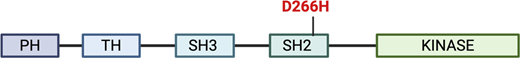
Structure of the ITK kinase and mutation.
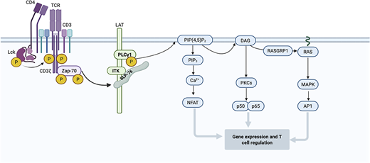
ITK signaling.
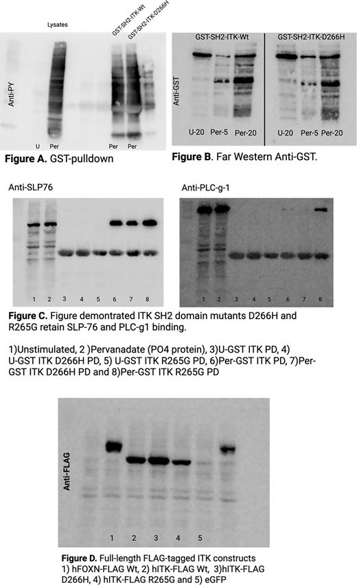
Results.


