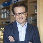
Members of the editorial board play a critical role at JCB. Our board members help authors navigate the editorial process by actively engaging with peer reviewers, offering guidance during revision, and ultimately determining the work that ends up in the pages of the journal. To best serve our authors, our board must therefore reflect the expanding multidisciplinary cell biology community. Thus, we are extremely pleased to welcome 24 new JCB editorial board members from across the breadth of cell biology. These scientists bring new voices to the journal and offer significant expertise across our ever-growing field. We welcome our new members, and are grateful to all of our editorial board members for their contributions to JCB and service to the community.
Laura Attardi
Cancer biology
Laura D. Attardi received her BA from Cornell University and her PhD from the University of California, Berkeley, where she worked with Professor Robert Tjian. Dr. Attardi did her postdoctoral training with Professor Tyler Jacks at the Massachusetts Institute of Technology Center for Cancer Research. In 2000, Dr. Attardi joined the Departments of Radiation Oncology and Genetics at the Stanford University School of Medicine, and she was promoted to professor in 2014. The focus of her laboratory is to dissect the pathways by which p53 acts in vivo using the mouse as a model system. She has been a recipient of a Damon Runyon Scholar Award, an American Cancer Society Research Scholar Grant, and a Leukemia and Lymphoma Society Scholar Award. She was named an American Association for the Advancement of Science fellow in 2007. In 2015, she received the National Cancer Institute Outstanding Investigator Award. Photo courtesy of Karin Kao.
Johan Auwerx
Integrative systems physiology
Johan Auwerx is a professor at the École Polytechnique Fédérale in Lausanne, Switzerland. Dr. Auwerx has been using molecular physiology and systems genetics to understand mitochondrial function and organismal metabolism in health, aging, and disease. Much of his work has focused on understanding how diet, exercise, and hormones control mitochondrial metabolism through changing the expression of genes by altering the activity of transcription factors and their associated cofactors. His work was instrumental for the development of several compounds that are currently used to treat high blood lipid levels, fatty liver, and type 2 diabetes. He was elected as a member of the European Molecular Biology Organization in 2003 and has received many international scientific prizes. Dr. Auwerx received both his MD and PhD in molecular endocrinology at the Katholieke Universiteit in Leuven, Belgium. He was a postdoctoral research fellow in the Departments of Medicine and Genetics of the University of Washington in Seattle. Photo courtesy of the École Polytechnique Fédérale de Lausanne.
Bill Bement
Signal transduction, cytoskeleton, and intracellular pattern formation
Bill Bement is a professor of zoology and the director of the Laboratory of Cell and Molecular Biology at the University of Wisconsin-Madison. Dr. Bement earned a BA in biology from Whitman College, earned a PhD in zoology/cell and developmental biology at Arizona State University under the direction of David Capco, and conducted his postdoctoral research in cell and molecular biology in the laboratory of Mark Mooseker at Yale University. The research in Dr. Bement’s lab is focused on understanding how cells link and integrate signals to control complex cellular processes based on the cytoskeleton, including cell repair and cell division. Photo courtesy of L.D. Bronstein, University of Wisconsin-Madison.
Sue Biggins
Chromosome segregation and cell cycle control
Sue Biggins studies the mechanisms that ensure accurate chromosome segregation and regulation of the cell cycle. Dr. Biggins obtained her PhD in molecular biology from Princeton University and went on to do postdoctoral work at the University of California, San Francisco, in Dr. Andrew Murray’s lab. She joined the faculty in the Division of Basic Sciences at the Fred Hutchinson Cancer Research Center in 2000, where she is currently a full member and associate director, as well as an investigator of the Howard Hughes Medical Institute. Her research interests surround the mechanisms that regulate chromosome segregation and the spindle checkpoint. Her lab studies the specialized chromatin that occurs at centromeres and demonstrated that a single nucleosome exists at the budding yeast centromere. Recently, her lab achieved the first isolation of native kinetochores and is currently applying structural, biophysical, and biochemical techniques to elucidate the mechanisms of kinetochore–microtubule interactions and spindle checkpoint regulation. Photo courtesy of Ron Wurzer.
Julius Brennecke
Small RNA silencing pathways in genome defense
Julius Brennecke is a senior investigator at the Institute for Molecular Biotechnology of the Austrian Academy of Sciences (IMBA) in Vienna. After studying biology at the University of Heidelberg (Germany) he joined the group of Steve Cohen at the European Molecular Biology Laboratory (EMBL) for his PhD training. There, he became interested in small RNA silencing pathways and studied the biology of microRNAs in Drosophila. For his postdoctoral studies, he moved to Cold Spring Harbor Laboratories and worked with Greg Hannon on the gonad-specific Piwi–piRNA pathway that silences transposable elements in animal germ cells. In 2009, Dr. Brennecke started his independent research group at IMBA, an institute dedicated to basic research in molecular biology. His team studies the ancient arms race between transposons and the host genome in gonads with a focus on Drosophila ovaries. Here, the Piwi–piRNA pathway acts as a small RNA based genome immune system by suppressing the deleterious activity of selfish genetic elements during gametogenesis. Photo courtesy of Julius Brennecke.
Nika N. Danial
Cellular fuel preferences and metabolic control of cell fate and function
Nika Danial is an associate professor of cell biology at the Dana-Farber Cancer Institute, Harvard Medical School. She obtained her PhD from Columbia University, and subsequently trained with the late Stanley J. Korsmeyer as a postdoctoral fellow at the Dana-Farber Cancer Institute, where she identified a novel role for the BCL-2 family protein BAD in glucose metabolism independent of its apoptotic function. The broad goal of the research in her laboratory is to determine how alternate fuel sources (carbohydrates, fatty acids, and ketone bodies) modulate cellular stress responses. Toward this goal, her group uses contemporary tools of mouse genetics, chemical biology, proteomics, and metabolomics to determine both the molecular mechanisms and biologic consequences of reprogramming cellular fuel preferences. This line of investigation has provided molecular insights into cellular adaptation to nutrient availability, metabolic control of seizure responses, and metabolic heterogeneity in tumors. Photo courtesy of Nika N. Danial.
Jan Ellenberg
Cell division and nuclear biogenesis
Jan Ellenberg heads the Cell Biology and Biophysics Unit at the EMBL in Heidelberg, Germany. Over the past 20 years Dr. Ellenberg has been interested in cell division and nuclear biogenesis, including systems analysis of mitosis, nuclear pore complex assembly, and formation of mitotic chromosomes. His goal has been to obtain structural and functional measures of the required molecular machinery inside cells using quantitative 4D imaging, single molecule spectroscopy, as well as super-resolution microscopy, which his group is constantly automating to address all molecular components comprehensively. His research group played a key role in large EU-wide efforts in microscopy automation and unbiased computational image analysis (http://www.mitocheck.org, http://www.mitosys.org, and http://www.systemsmicroscopy.eu) establishing methods to reliably score up to billions of cells and capture rare and transient functional states automatically. Because of the important role of new imaging technologies for the future of the life sciences, he has coordinated European efforts to make imaging technologies more accessible to researchers as open access research infrastructures, which he continues to promote in his role as EMBL delegate of Euro-BioImaging (http://www.eurobioimaging.eu). Photo courtesy of EMBL.
Marc Freeman
Neuron-glia signaling
Marc Freeman started his laboratory at The University of Massachusetts Medical School in 2004 (Worcester, MA). The Freeman laboratory studies a number of topics relevant to neuron-glia signaling in the healthy and diseased brain, including glial engulfment of neuronal cell corpses and axonal debris, molecular mechanisms of axon auto-destruction, and mechanisms by which glia control neural circuit assembly and function. The model systems they use for their studies include Drosophila, mice, and human cell lines. Freeman is a professor and vice chairman of the Department of Neurobiology, and in 2013 was appointed an investigator of the Howard Hughes Medical Institute. Photo courtesy of Marc Freeman.
Melissa Gardner
Biophysical studies of mitotic microtubule dynamics and spindle function
Melissa Gardner is an assistant professor at the University of Minnesota, Department of Genetics, Cell Biology, and Development. She received her PhD in biomedical engineering from the University of Minnesota and completed postdoctoral work in biophysics with Joe Howard at the Max Planck Institute of Molecular Cell Biology and Genetics in Dresden, Germany. Her research group uses a combination of experimental and computational approaches to dissect molecular mechanisms for how cells divide, and for how cell division can be controlled to prevent genetic diseases, pathogenic anti-fungal drug resistance, and cancer. Specifically, her research team in interested in how proteins regulate the dynamics and mechanics of critical cell division components such as microtubules and chromosomes, with the overarching goal of improving human health by developing an improved mechanistic understanding for how cells divide. Photo courtesy of the University of Minnesota.
Johanna Ivaska
Cell adhesion and cancer
Johanna Ivaska is academy professor and professor of molecular cell biology at the University of Turku in Finland. Her research group studies the biological role of integrins in cancer progression. Her current main research focus areas are integrin-mediated cell adhesion, cell–matrix interactions, and integrin endosomal traffic in cancer. Johanna completed her PhD at the University of Turku with Jyrki Heino, where she studied collagen-binding integrins. She then did her postdoctoral training with Peter J. Parker at the Cancer Research UK, London Research Institute, working on protein kinase C and integrin endosomal trafficking. She was academy research fellow from 2003 and professor from 2008 at VTT Technical Research Centre of Finland until she moved to the University of Turku Centre for Biotechnology in 2013. To find out more please visit the Ivaska lab websites: http://www.ivaskalab.com and http://www.btk.fi/research/research-groups/ivaska/. Photo courtesy of Hanna Oksanen, University of Turku, Finland.
Susan Kaech
Immune cell development and homeostasis
Susan Kaech is a professor of immunobiology at Yale University School of Medicine. After completing her PhD in developmental biology with Stuart Kim at Stanford University, Susan moved to Emory University to study the development of memory CD8 T cells with Rafi Ahmed. Since moving to Yale in 2004, Susan has continued to investigate the mechanisms that regulate the generation and survival of memory T cells in response to both infection and vaccination. Her lab currently focuses on the transcriptional, epigenetic, and metabolic responses of T cells to signals in the tissue microenvironment, and also how these cells recognize and respond to tumors. Susan was a Howard Hughes Medical Institute Early Career Scientist and has received a Presidential Early Career Award for Scientists and Engineers, as well as investigator awards from the Cancer Research Institute and American Asthma Foundation. Photo courtesy of Yale University School of Medicine.
Scott Keeney
Meiotic recombination
Scott Keeney is a member of the Molecular Biology Program, Memorial Sloan Kettering Cancer Center (MSKCC), and Howard Hughes Medical Institute investigator. He is also a professor in the Louis V. Gerstner Graduate School of Biomedical Sciences (MSKCC) and the Weill Cornell Graduate School of Medical Sciences. Scott received his PhD in biochemistry from the University of California, Berkeley, and completed postdoctoral work at Harvard University. Research in his lab aims to understand the mechanism and regulation of homologous recombination during meiosis. His group studies meiosis principally in mouse and in S. cerevisiae, using molecular genetic, biochemical, genomic, and cytological approaches. As a postdoc, he discovered that Spo11 is the protein that generates the double-strand breaks that initiate meiotic recombination. As an independent researcher, his lab discovered a pathway for endonucleolytic processing of covalent protein-linked double-strand breaks; devised novel strategies for mapping the genome-wide distribution of breaks; demonstrated a mechanism that maintains crossover numbers when double-strand break numbers are decreased (a phenomenon we call crossover homeostasis), and dissected negative feedback pathways that control break numbers. Photo courtesy of Marsha Henderson.
Juergen Knoblich
Stem cells and asymmetric cell division
Juergen Knoblich is a senior scientist and deputy director at IMBA, Vienna. He obtained his PhD from the Max Planck Institute in Tübingen. After a postdoctoral period in the laboratory of Yuh Nung Jan at the University of California, San Francisco, he joined the Institute of Molecular Pathology in 1997 as a junior group leader. In 2004, he moved to IMBA, where he is now senior scientist and deputy director. His laboratory is interested in the biology of neural stem cells. In the fruit fly, they have identified the molecular mechanism that allows neural stem cells to divide asymmetrically and segregate protein determinants into only one daughter cell during mitosis. They have demonstrated that defects in this mechanism lead to brain tumor formation. More recently, they have extended their interest to analyzing mammalian neural progenitors and their contribution to brain development. To analyze those processes in humans, they have established a 3D culture system that recapitulates the early steps of human brain development in cell culture allowing brain pathologies and human-specific developmental events to be studied in unprecedented detail. Photo courtesy of IMBA.
Alex Mogilner
Cell motility, mitosis, galvanotaxis, and modeling in cell biology
Alex Mogilner is a professor of mathematics and biology at the Courant Institute and in the Department of Biology at New York University. Dr. Moligner received his PhD in applied mathematics from the University of British Columbia and completed his postdoctoral work in computational and cell biology at the University of California, Berkeley. His laboratory uses methods of mathematical and computational modeling, together with experimental data analysis, to understand mechanics of cell migration, mitosis, and galvanotaxis. Dr. Mogilner proposed an elastic polymerization ratchet model explaining cell protrusions, models of graded actomyosin contraction, and actin treadmilling array in a membrane “bag” that predict motile cell shapes and speeds. Together with experimentalists, he elucidated feedback mechanisms underlying cell polarization and motility initiation. He participated in developing and testing a force-balance model of the mitotic spindle and search-and-capture model of spindle assembly. Photo courtesy of Cynthia Lee, Mechonobiology Institute, National University of Singapore, Singapore.
Karla Neugebauer
RNA biology
Karla Neugebauer is professor of molecular biophysics and biochemistry at Yale University. Her lab investigates roles for RNA in the organization and function of living cells, focusing on: (1) coordination between RNA processing, transcription, and chromatin and (2) RNA-rich cellular subcompartments, such as Cajal bodies, that facilitate RNA processing and ribonucleoprotein assembly. Her lab discovered transcriptional pausing associated with co-transcriptional splicing in budding yeast and feedback from splicing to transcription and chromatin in human cells. Her lab developed the zebrafish embryo as a model system for studying nuclear organization, showing that snRNP assembly in Cajal bodies is essential for embryogenesis. She obtained her PhD in neuroscience at University of California, San Francisco, working with Louis Reichardt on cell adhesion and axon growth. As a postdoctoral fellow with Mark Roth at the Fred Hutchinson Cancer Research Center, she became fascinated by RNA. She was research group leader at the Max Planck Institute of Molecular Cell Biology and Genetics in Dresden, Germany, from 2001 to 2013. Photo courtesy of William Sacco.
Eva Nogales
Visualization of macromolecular structure and function
Eva Nogales is a professor in the Molecular and Cell Biology Department at the University of California, Berkeley, a Howard Hughes Medical Institute investigator, and a senior faculty scientist at Lawrence Berkeley National Laboratory (LBNL). She received her BA in physics from the Universidad Autónoma de Madrid. She carried out her graduate work in biophysics at the Synchrotron Radiation Source in the UK. Her postdoctoral research at LBNL in the lab of Ken Downing resulted in the electron crystallographic structure of tubulin. Her lab is dedicated to gaining mechanistic insight into two important areas of eukaryotic biology: central dogma machinery in the control of gene expression and cytoskeleton interactions and dynamics in cell division. She uses state-of-the-art cryoelectron microscopy and image analysis, as well as biochemical and biophysical assays. Her most recent studies include the atomic structure of microtubules in different functional states and the structural characterization of the assembly and architecture of the human transcription preinitiation complex. Photo courtesy of Amparo Garrido, Centro Nacional de Investigaciones Oncológicas, Madrid, Spain.
Ana Pombo
Epigenetic regulation and chromatin architecture
Ana Pombo is a group leader at the Berlin Institute for Medical Systems Biology (BIMSB) of the Max Delbrueck Center for Molecular Medicine (MDC) and a professor (W3) at Humboldt University, Berlin, Germany. She received her PhD from the University of Oxford, UK, working on transcription factories with Peter Cook, after which she was awarded the Hayward Fellowship (Oriel College, Oxford, UK) and the Royal Society Dorothy Hodgkin Fellowship (UK). In 2000, she established her independent research group at the Medical Research Council Clinical Sciences Centre in London, working on gene expression and genome architecture, and from 2013 at BIMSB, MDC. In 2007, she received the Robert Feulgen Prize for her contributions to imaging nuclear architecture. Her laboratory currently studies how the 3D folding of chromatin influences gene expression in development and disease, and the mechanisms of gene priming and activation in ES and differentiated cells, through posttranslational modifications of RNA polymerase II C-terminal domain. Photo courtesy of David Ausserhofer, © MDC.
Craig Roy
Microbial pathogenesis
Craig Roy is a professor and vice-chair of the Department of Microbial Pathogenesis at Yale University School of Medicine. Craig received his PhD in microbiology and immunology from Stanford University and completed a postdoctoral fellowship at Tufts University in Boston. Research in his laboratory focuses on the cell biology of bacterial infection using Legionella pneumophila and Coxiella burnetii as model pathogens. His group has discovered novel proteins that intracellular pathogens use to modulate specific host membrane transport pathways, which allow these pathogens to evade cell autonomous defenses and create novel organelles that permit bacterial replication. Photo courtesy of Terry Dagradi, Yale University.
Andrey Shaw
Podocyte biology and immune cell signal transduction
Andrey Shaw recently joined Genentech as a staff scientist. Previously, he was the Emil R. Unanue Professor and head of the Division of Immunobiology at Washington University as well as an investigator of the Howard Hughes Medical Institute. Dr. Shaw received his BA and MD degrees from Columbia University and completed pathology residency training at Yale University. His postdoctoral training was in cell biology at Yale with Jack Rose. His work is generally focused on signal transduction mechanisms used by immune cells and understanding the role of kidney podocytes in renal filtration. His current work focuses on kinase regulation as well as using a variety of imaging and computational approaches to understand the signaling of immune cells in vivo and the kidney podocyte. Photo courtesy of Andrey Shaw.
Zu-Hang Sheng
Axonal transport of mitochondria, endolysosomes, and synaptic cargoes
Zu-Hang Sheng is a senior investigator and chief of the Synaptic Function Section at the National Institute of Neurological Disorders and Stroke, National Institutes of Health, in Bethesda, MD. He received his PhD in biochemistry from the University of Pennsylvania with Roland Kallen and Robert Barchi. He completed his postdoctoral research with William Catterall at the University of Washington. His laboratory focuses on mechanisms regulating axonal transport that are essential for the maintenance of synaptic function and axonal homeostasis. Using genetic mouse models, his group is addressing several fundamental questions: (1) how mitochondrial transport is regulated to sense changes in synaptic activity, mitochondrial integrity, axon injury, and pathological stress; (2) how neurons coordinate late endocytic transport and autophagy-lysosomal function to maintain cellular homeostasis; and (3) how impaired transport contributes to synaptic dysfunction and axonal pathology in neurodegenerative diseases. These studies have led to the identification of several motor adaptor and anchoring proteins that regulate axonal transport of mitochondria, endolysosomes, and synaptic cargos. Photo courtesy of the National Institute of Neurological Disorders and Stroke, National Institutes of Health.
Michael Sixt
Morphodynamics of immune cells
Michael Sixt is a professor at the Institute of Science and Technology Austria (IST Austria), where he studies the mechanistic principles underlying cellular locomotion. His group focuses mainly on rapidly migrating immune cells and explores how these cells generate intracellular force, how they transduce the force to the environment, and how cells are directionally guided within the tissue context. Dr. Sixt trained as a medical doctor at the University of Erlangen in Germany, where he also worked as a clinical resident in dermatology. After finishing his medical dissertation on the role of basement membranes in leukocyte extravasation he was a postdoc in the laboratory of Lydia Sorokin in Lund, Sweden, where he explored microanatomical features of lymphatic organs. Subsequently, he established his own lab at the Max Planck Institute of Biochemistry in Martinsried, Germany, where he started to investigate cell biological and biophysical aspects of migrating leukocytes. In 2010, he moved to the then newly established IST Austria. Photo courtesy of IST Austria.
Aaron Straight
Chromosome structure and function
Aaron Straight is an associate professor in the Biochemistry Department at Stanford University. His PhD training was with Andrew W. Murray at the University of California, San Francisco, studying the process of chromosome segregation. He went on to join Tim Mitchison’s group in the Department of Cell Biology and the Institute for Chemistry and Cell Biology at Harvard Medical School studying cytokinesis. His research at Stanford has focused on chromosome segregation mechanisms and, in particular, the formation and function of centromeres. Using diverse model organisms and approaches, his group has developed new systems to understand, at a mechanistic level, the epigenetic control of centromere establishment and assembly. Current efforts in his group are exploring the multiscale organization of eukaryotic genomes, the control of zygotic genome activation, and the regulation of heterochromatin formation. Photo courtesy of Whitney Johnson.
Erwin F. Wagner
Genes, development, and disease
Erwin Wagner received his university education in Austria and obtained his PhD in 1978 for studies performed in Berlin on bacterial genetics. He joined the lab of Beatrice Mintz in Philadelphia in 1979 for his postdoctoral training, where he developed microinjection of DNA into fertilized eggs and gene transfer technologies into mice. In 1983 he became group leader at the EMBL in Heidelberg and, in 1988, joined the IMP in Vienna as a founding member. He became professor of the University of Vienna in 1994 and deputy director at the IMP. In 2008 he moved to the Centro Nacional de Investigaciones Oncológicas in Madrid, as vice director and director of the cancer cell biology program. His work focuses on the functions of the AP-1 (Fos/Jun) transcription factor complex, e.g., in inflammation and cancer, using the mouse as a model organism and human patient samples. He also developed mouse models for common human diseases such as osteoporosis, fibrosis, and psoriasis, to be applied for preclinical studies. Photo courtesy of Erwin F. Wagner.
Tobias Walther
Lipid and membrane homeostasis
Dr. Walther studies organelle, membrane, and lipid homeostasis. He completed his PhD at the EMBL and postdoctoral studies at the University of California, San Francisco. After that, Dr. Walther led a laboratory at the Max Planck Institute of Biochemistry and Yale School of Medicine. At Harvard since 2014, Dr. Walther and his scientific partner Robert Farese Jr. study how cells perceive changes in membrane lipids and regulate metabolism and cell biology accordingly. The laboratory studies mechanisms of neutral lipid storage and the cell biology of cytosolic lipid droplets. In addition, the Farese and Walther laboratory studies homeostasis mechanisms for lipids not stored in lipid droplets and how they contribute to physiology and disease. Dr. Walther is an investigator of the Howard Hughes Medical Institute, a professor of genetics and complex diseases (Harvard T.H. Chan School of Public Health) and cell biology (Harvard Medical School) at Harvard University, and an associate member of the Broad Institute of the Massachusetts Institute of Technology and Harvard. Photo courtesy of Aynsley Floyd, Associated Press.


